
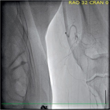
Figure 1.
Angiogram showing the acute vascular occlusion at the left brachial artery.
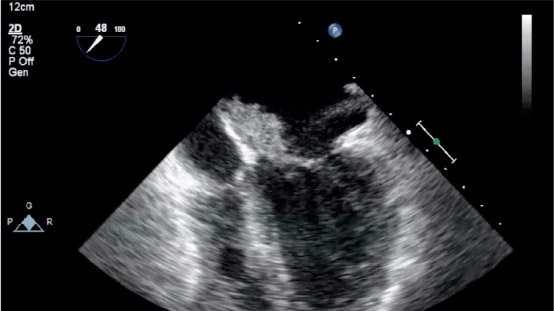
Figure 2.
Transesophageal echocardiography revealing myxoma on left atrium.
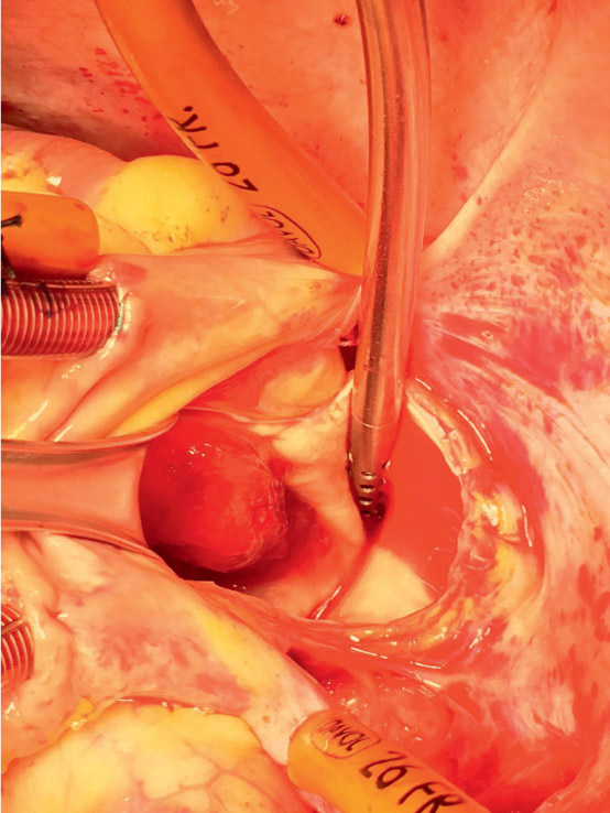
Figure 3.
Cardiac myxoma in the left atrium.
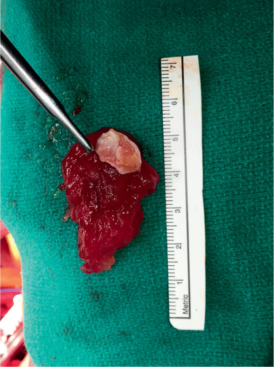
Figure 4.
The excised cardiac myxoma: soft gelatinous-like with irregular fronds.
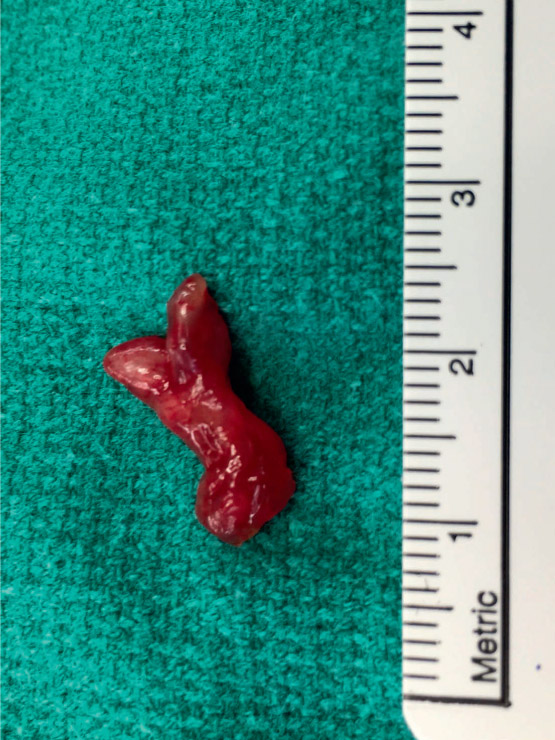
Figure 5.
Tumor embolus removed from the brachial artery.