
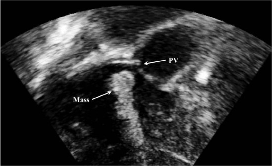
Figure 1.
Transesophageal echocardiogram reveals an echodense structure in the right ventricular outflow tract (arrow). PV: pulmonary valve.
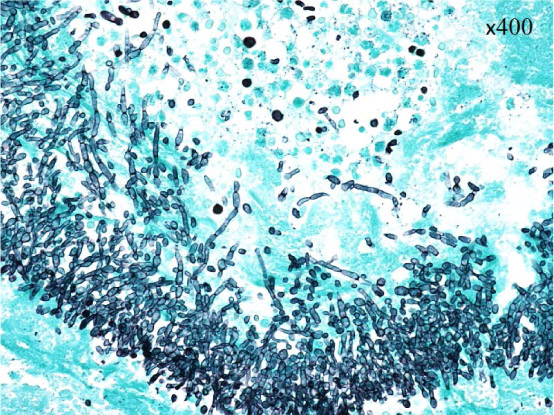
Figure 2.
Photomicrograph of the resected right ventricular mass revealing numerous budding yeast and pseudohyphae (GMS × 400).