
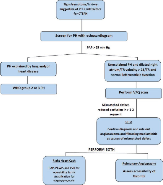
Figure 1.
Diagnostic algorithm for chronic thromboembolic pulmonary hypertension. PH: pulmonary hypertension; CTEPH: chronic thromboembolic pulmonary hypertension; TR: tricuspid regurgitation; WHO: World Health Organization; V/Q: ventilation/perfusion; PAP: pulmonary artery pressure; PCWP: pulmonary capillary wedge pressure; PVR: pulmonary vascular resistance; CTPA: computed tomography pulmonary angiogram.
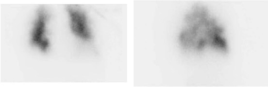
Figure 2.
Ventilation/perfusion scans with marked heterogeneity of lung perfusion bilaterally, highly suspicious of CTEPH.
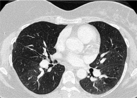
Figure 3.
Chest CT angiogram demonstrating marked mosaic attenuation in bilateral lung fields as well as pruning of distal pulmonary arteries and enlargement of proximal pulmonary arteries.
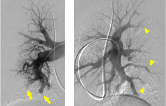
Figure 4.
Digital subtraction pulmonary angiography performed in anterior-posterior and lateral views of the right pulmonary artery demonstrating severe subsegmental disease. Multiple sites of abrupt narrowing (arrowhead) and pouch defect and subtotal occlusions (arrow) are visible.
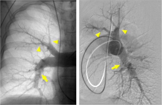
Figure 5.
Digital subtraction and native pulmonary angiography performed in anterior-posterior and lateral views of the right pulmonary artery demonstrating severe segmental disease. There are multiple sites of pruning and abrupt narrowing (arrowhead). The hazy band across the right interlobar artery is suggestive of a web lesion (arrow).
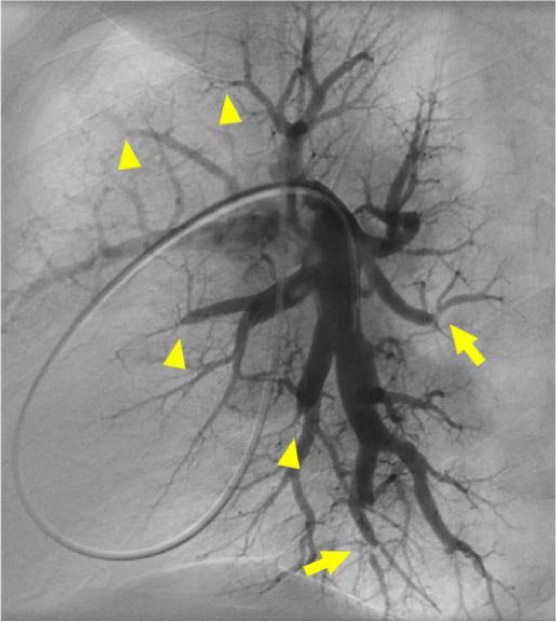
Figure 6.
Digital subtraction pulmonary angiography, lateral view of the left pulmonary artery demonstrating severe subsegmental disease. Multiple sites of subtotal and total occlusions (arrowhead) are visible. There are also multiple severe ring lesions (arrow).