
Have a personal or library account? Click to login
Position-Dependent Ventricular Tachycardia Related to Peripherally Inserted Central Venous Catheter Cover

Position-Dependent Ventricular Tachycardia Related to Peripherally Inserted Central Venous Catheter

Open Access
|Jul 2016

Figures & Tables

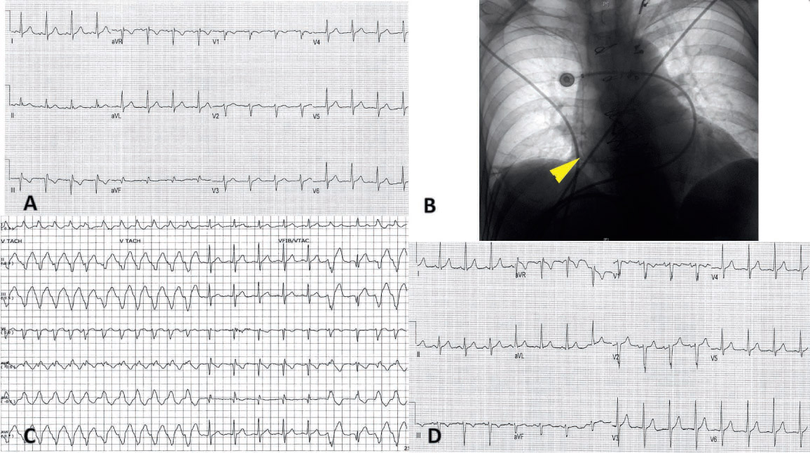
Figure 1.

(A) Baseline electrocardiogram (ECG) of patient. (B) Chest X-ray after initial peripherally inserted central catheter (PICC) line placement. Course of PICC line to floor of right atrium can be seen (yellow arrowhead). (C) Telemetry strip of runs of mechanical induction of nonsustained ventricular tachycardia by PICC line during patient positioning. (D) Sinus rhythm ECG after withdrawal of PICC line to superior vena cava-right atrium junction.

Language: English
Page range: 177 - 178
Published on: Jul 1, 2016
Published by: Houston Methodist DeBakey Heart & Vascular Center
In partnership with: Paradigm Publishing Services

© 2016 Paulino Alvarez, Paul Schurmann, Melanie Smith, Miguel Valderrábano, C. Huie Lin, published by Houston Methodist DeBakey Heart & Vascular Center
This work is licensed under the Creative Commons Attribution-NonCommercial 4.0 License.