
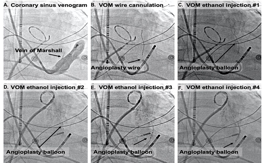
Figure 1.
Procedural steps for atrial fibrillation ablation using Vein of Marshall (VOM) ethanol infusion. (A) Coronary sinus (CS) venogram. From the internal jugular venous access, the CS is engaged with a sheath. Inside the CS sheath, a sub-selector left internal mammary artery (LIMA) guide catheter is inserted, and contrast is injected with the LIMA catheter tip pointing superiorly. The VOM is clearly visible via a nonselective injection through the LIMA. (B) Preloaded angioplasty wire and balloon are advanced into the VOM. Once the wire is securely engaged, the balloon is advanced as distally as possible in the VOM. (C-F) Balloon locations for the (C) first, (D) second, (E) third, and (F) fourth injection of ethanol in the VOM. Some myocardial staining occurs when contrast is injected after ethanol.
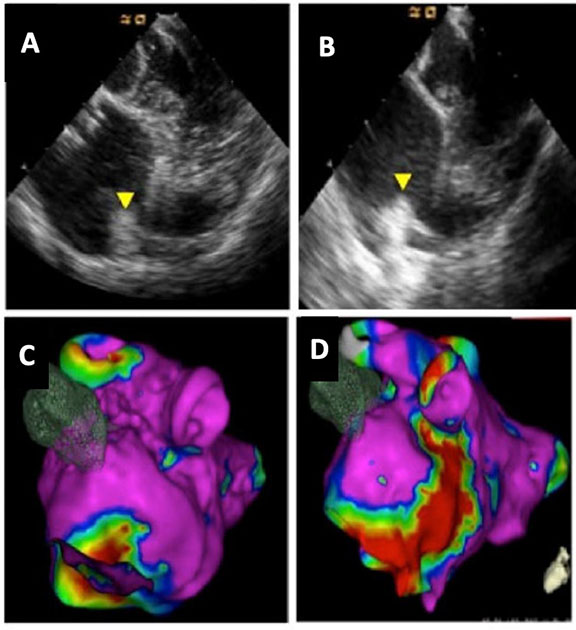
Figure 2.
Pre- and post-ethanol imaging demonstrating the scar created by Veinof Marshall (VOM) ethanol infusion using intracardiac echocardiogramimaging of (A, B) the left atrial ridge and (C, D) electroanatomical mapping.8
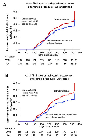
Figure 3.
Data from the VENUS Clinical trial. Time to recurrence of atrial fibrillationor tachycardia (A) as randomized and (B) as treated analysis.20