

Figure 1.
Top row: Preprocedural computed tomography imaging showing a complete “twist” of the outflow graft causing left ventricular assist device obstruction. Bottom row: Postprocedural imaging shows device exchange without twisting.
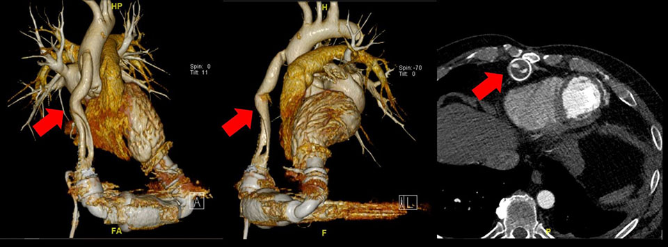
Figure 2.
Chest computed tomography showing proximal obstruction of the outflow tract.

Figure 3.
Intraoperative photo of external compression of the graft from old, dark, thrombotic gelatinous material within the bend relief without evidence of graft twist.