

Figure 1.
Explanted porcine heart after bipolar clamp application externally on the beating heart. Transmural lesions are seen on the endocardium. Reprinted with permission from Ann Cardiothorac Surg, 2014.19

Figure 2.
Left superior and inferior pulmonary veins are visualized, and the dry bipolar clamp is seen on the left antrum. Reprinted with permission from Ann Cardiothorac Surg, 2014.19

Figure 3.
Right-sided video-assisted thoracoscopic approach with a scope in the inferior port and working port being created in the anterior fourth intercostal space. Reprinted with permission from Ann Cardiothorac Surg, 2014.16

Figure 4.
The septal lesion with a bipolar clamp encompassing the right side of the left antrum as well as a Waterston groove fat pad; this extends from the base of the superior vena cava superiorly to the base of the inferior vena cava inferiorly. To the right is a bipolar pacing lead inside of the pericardium. Reprinted with permission from Ann Cardiothorac Surg, 2014.16

Figure 5.
The stapled left atrial appendage is being removed from the intrapericardial area. Reprinted with permission from Ann Cardiothorac Surg, 2014.16

Figure 6.
Intraoperative photograph demonstrating the position of the camera port with a chest tube and a 6-cm working port in the fourth intercostal space anteriorly. Reprinted with permission from Ann Cardiothorac Surg, 2014.16

Figure 7.
The usual testing areas for ganglionic plexi (GP) activity on the (A) right side and (B) left side of the left atrium.19 SVC: superior vena cava; RSPV: right superior pulmonary vein; RIPV: right inferior pulmonary vein; LA: left atrium; RA: right atrium; IVC: inferior vena cava; LAA: left atrial appendage; SLPV: left superior pulmonary vein; LV: left ventricle; LIPV: left inferior pulmonary vein; AV groove: auriculoventricular groove
Table 1.
Different tests used during treatment of lone atrial fibrillation.
| Imaging/Test | Purpose |
|---|---|
| Electrocardiograph | Document rhythm |
| Echocardiogram | Measure size of left atrium; confirm valve function |
| 64-slice computed tomography | Visualize anatomy of pulmonary veins (PV) and left atrial appendage; identify possible thrombus, PV stenosis, coronary anatomy pathology |
| Home monitoring (if needed) | Document atrial fibrillation burden |

Figure 8.
Minimally invasive left-sided Cox Maze 4. Through a rightthoracotomy, on bypass and open heart, a sequence of leftatrial endocardial cryo and radiofrequency ablations aredepicted. Reprinted with permission from AtriCure.

Figure 9.
For patients who cannot undergo a complete mini-maze procedure,a stand-alone minimally invasive technique using Atriclip left atrialappendage (LAA) closure allows consistent and complete closure of theLAA with excellent stroke prevention. In this operative photograph, thewhite Atriclip is seen at the base of the LAA.

Figure 10.
Sympathetic and parasympathetic innervation of the heart. Reprinted with permission from Springer Nature.31

Figure 11.
Active ganglionic plexi areas on the outside of the left atrium are shown inyellow.32 LIPV: left inferior pulmonary vein; LSPV: left superior pulmonaryvein; RSPV: right superior pulmonary vein; RIPV: right inferior pulmonaryvein; IVC: inferior vena cava
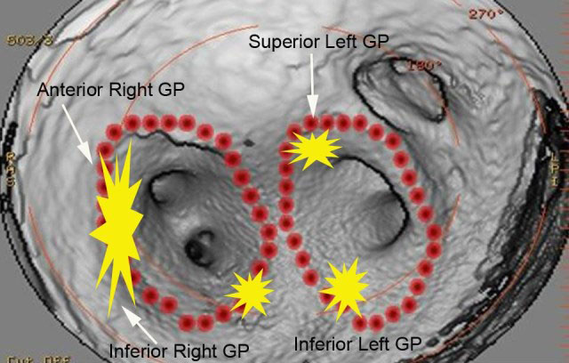
Figure 12.
Active ganglionic plexi areas are depicted in yellow. As shown on theinside of the left atrium, these areas are actually on the epicardialsurface. Reprinted with permission from Jeff Williams, MD, HeartRhythm Center blog. Image courtesy of Jeff Williams, MD.

Figure 13.
The CardioScout device includes (A) a tiny light source, camera, and(B) working ports for epicardial visualization of the beating heart.Images courtesy of James Fonger, MD