
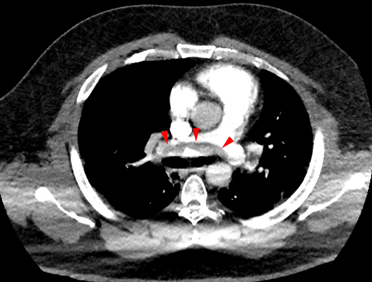
Figure 1
A large, saddle pulmonary embolism (arrows) shown in computed tomography angiogram of the chest, axial view.
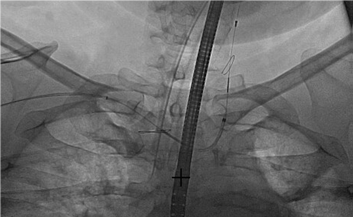
Figure 2
Fluoroscopic image of the SENTINEL cerebral embolic protection device (Boston Scientific) with proximal basket in the brachiocephalic trunk and distal basket in the proximal left carotid artery.
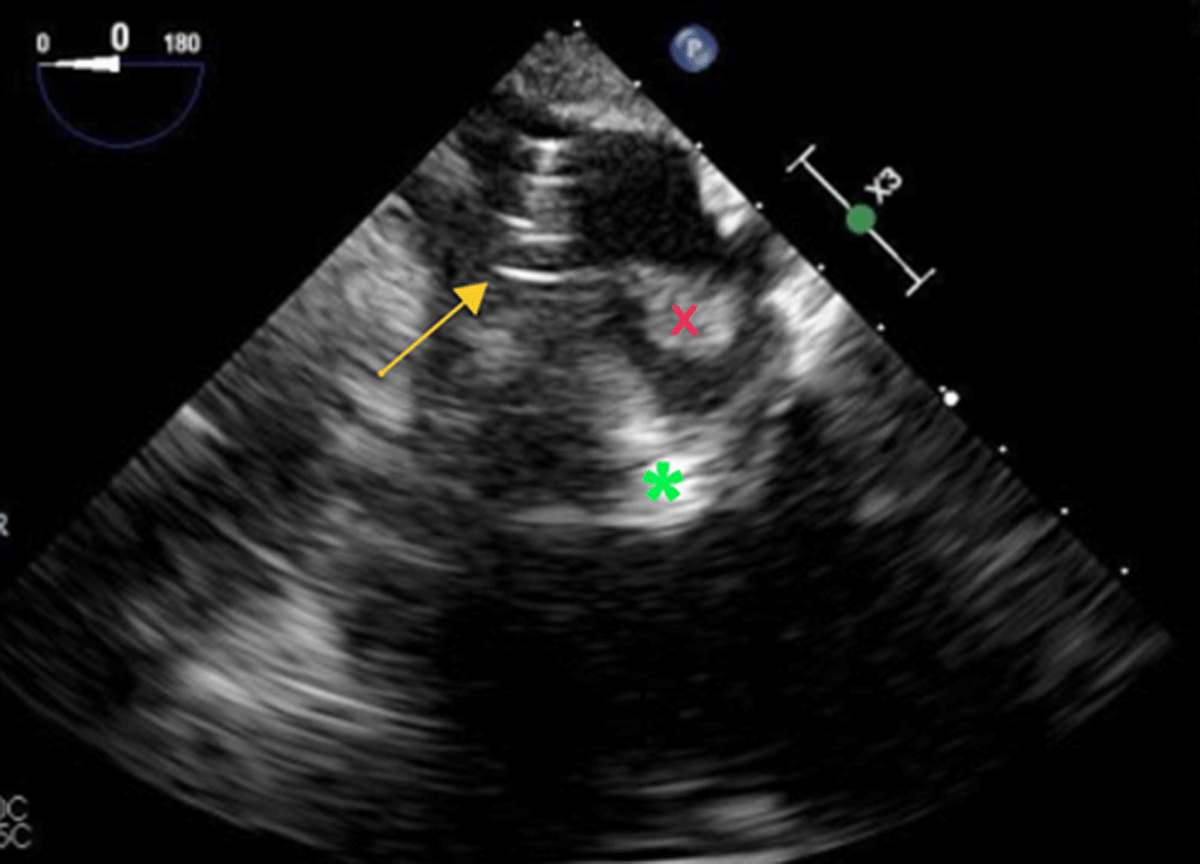
Figure 3
Transesophageal echocardiogram shows the AngioVac System cannula (arrow) retrieved back into the right atrium across the patent foramen ovale. The left atrial portion of the clot (X) is visualized, as is the right atrial portion of the clot (*).
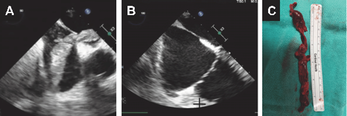
Figure 4
Transesophageal echocardiogram and clinical specimen. (A) Negative flow applied using the AngioVac system’s inflow aspirates the clots in a retrograde fashion into the right atrium across the patent foramen ovale. Microcavitation is seen on the right chamber and leftward interatrial septum. (B) Post clot extraction. (C) Extracted specimen.