
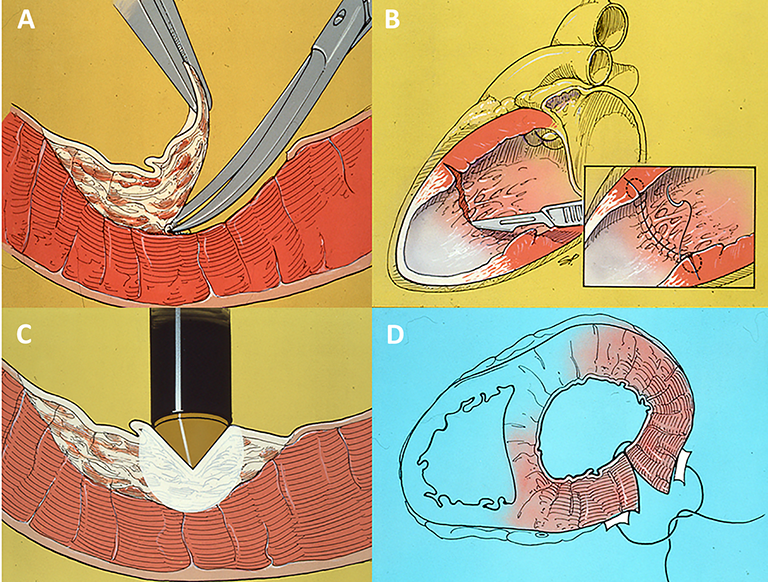
Figure 1.
Surgical techniques. (A) Endocardial resection. (B) Cryotherapy at -70°C. (C) Encircling endocardial incision. (D) Excision of arrhythmogenic tumors or myocardium.
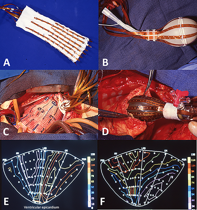
Figure 2.
(A) Epicardial mapping sock with Kapton strips holding electrodes. (B) Endocardial mapping balloon with Kapton strip electrodes. (C) Sock positioned on left ventricular (LV) epicardium, apex is to right. (D) Balloon being inserted in the beating heart through a small apical ventriculotomy into the LV, where it will be inflated. (E) Automated epicardial activation map in sinus rhythm. (F) Map in ventricular tachycardia showing inferoapical earliest activation below posteromedial papillary muscle.
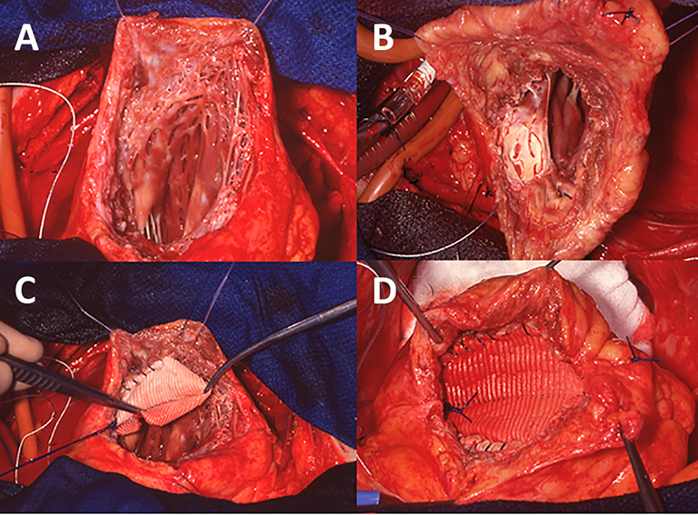
Figure 3.
Same patient as shown in Figure 2. (A) Anteroapical and inferior left ventricular aneurysm (LVA) opened and extensive endocardial resection performed. (B) Base of posteromedial papillary muscle cryoablated with 20 mm lesion. (C) Oval Dacron patch is being sewn to junction of LVA with normal tissue, deep within the fibrous LVA to remodel LV cavity. (D) Patch closure is completed. Scar of aneurysm will be closed over patch.
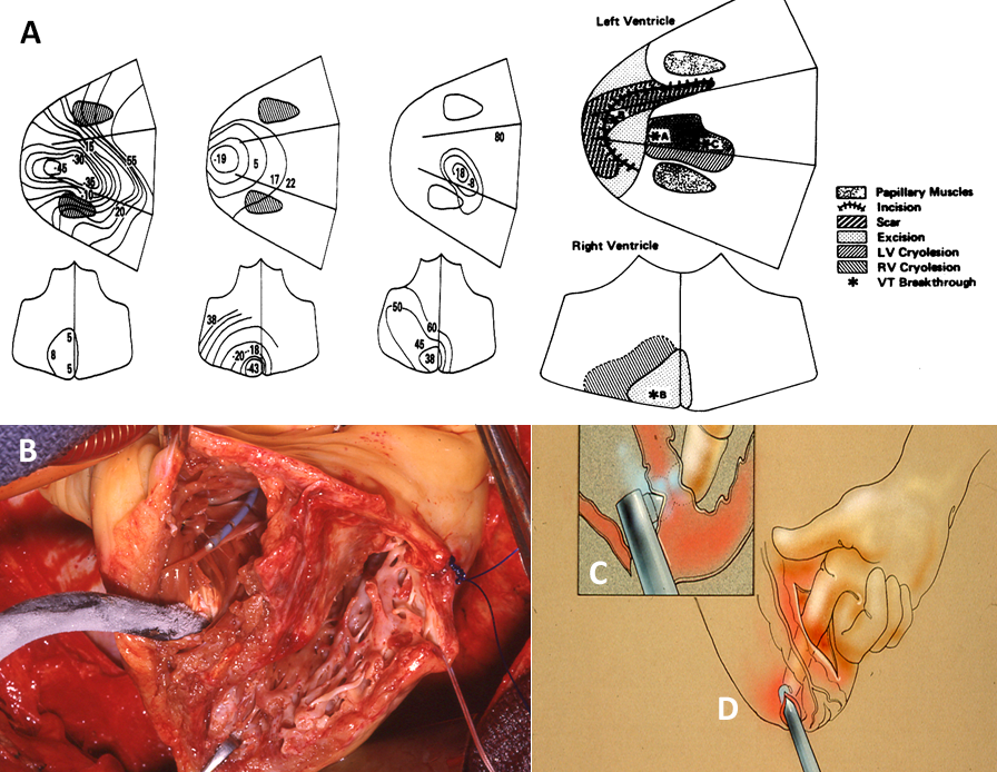
Figure 4.
(A) Patient with 3 ventricular tachycardia breakthroughs mapped pre- and intraoperatively, 2 on the left ventricular side of septum and 1 on the right ventricular side. Locations of breakthroughs and procedures performed are shown in diagram. (B) Biventricular cryotherapy was applied to the septum, C and D, as well as apical left ventricular aneurysm resection.
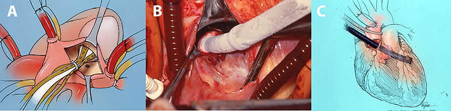
Figure 5.
Transannular balloon mapping and cryoablation of a high right ventricular septal nonischemic lesion.(A) Balloon inserted through tricuspid valve on the beating heart. (B) Transannular cryotherapy wasperformed on the cold arrested heart. No ventriculotomy was performed. (C) In several other cases,transaortic cryotherapy was performed.

Figure 6.
Governors' Award from American College of Cardiology (1983)