
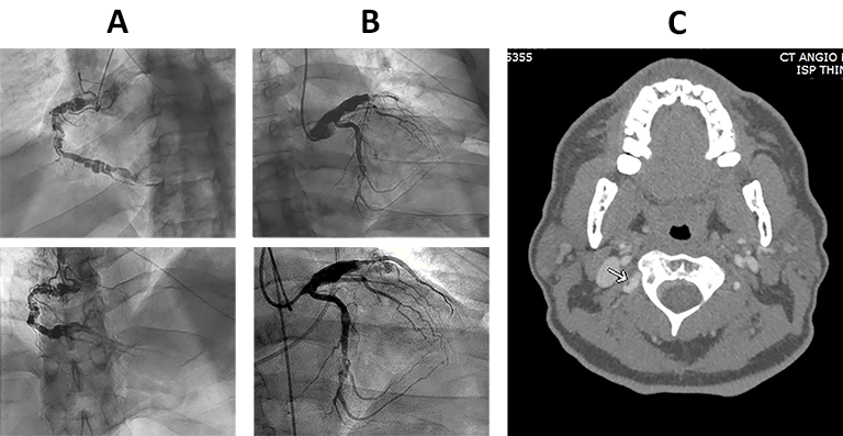
Figure 1.
(A) Coronary angiography of the right coronary artery demonstrating marked ectasia, critical narrowing, and aneurysmal dilatation. (B) Coronary angiography of the left coronary system demonstrating intraluminal filling defects in the left circumflex artery and the left anterior descending artery. (C) Representative computed tomography angiogram of the head demonstrating multifocal irregularity of the proximal V3 segment of the right vertebral artery.