
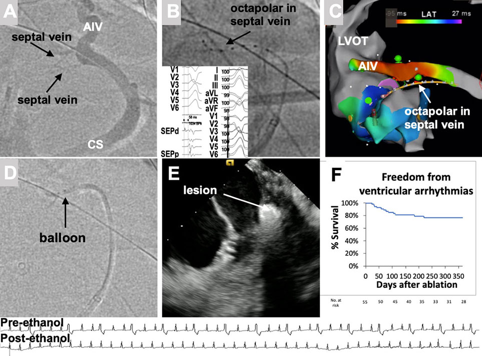
Figure 1.
Venous mapping and ethanol ablation technique. (A) Coronary sinus venogram (left anterior oblique caudal projection) showing two septal branches at the take-off of the anterior interventricular vein (AIV). (B) Octapolar catheter in the first septal vein. Intramural vein signals precede QRS by 58 ms, and pace-mapping reproduces exact QRS morphology (inset). (C) 3-dimensional activation map shows early (red) signals in the AIV and the location of the octopolar catheter. (D) Balloon cannulation of the septal vein. (E) Intramural echogenicity after venous ethanol, which leads to elimination of ectopy (bottom). (F) Kaplan-Meier plot of ablation success over 1-year follow-up in 55 patients with previous failed radiofrequency ablations. CS: coronary sinus; LVOT: left ventricle outflow tract
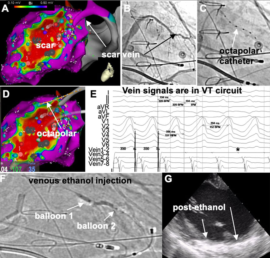
Figure 2.
Mapping and ethanol ablation of an infarct-related vein in ischemic ventricular tachycardia (VT). (A) Endocardial 3-dimensional bipolar voltage map of the left ventricle (LV) showing a dense (< 0.5 mV) scar, with a scar vein directly on its epicardial aspect. (B) Venography. (C) Octapolar catheter in the vein. (D) Localization of octapolar relative to scar. (E) Entrainment and mid-diastolic signals confirm participation in VT circuit. (F) Double balloon ethanol vein injection. (G) Transmural echogenicity in lateral LV after ethanol.