
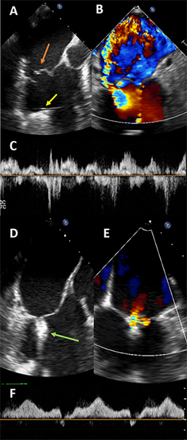
Figure 1.
Intraprocedural transesophageal echocardiogram images for patient 1. Panel A shows prolapse and flail with ruptured chordae of the A2 scallop (orange arrow). The left ventricular assist device inflow cannula is seen as well (yellow arrow). Panel B shows color Doppler across the mitral valve at Nyquist limit of 61 cm/second, suggesting severe mitral regurgitation (MR). Panel C shows a pulsed-wave (PW) Doppler through one pulmonary vein, showing systolic reversal, and was seen in all four pulmonary veins. After one MitraClip XTR was placed at the A2-P2 scallop (Panel D, green arrow), the severity of MR significantly decreased (panel E), and PW Doppler through the same pulmonary vein shows antegrade flow (Panel F) confirming mild MR. Mean gradient after clip placement is 2 mm Hg.
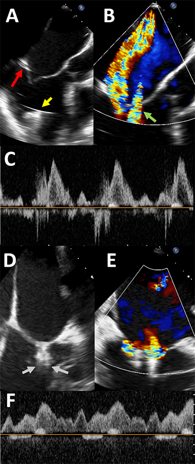
Figure 2.
Intraprocedural transesophageal echocardiogram images for patient 2. Panel A shows tethering of the posterior leaflet (red arrow) as well as the left ventricular assist device (LVAD) inflow cannula (yellow arrow). Panel B shows color Doppler across the mitral valve, confirming significant mitral regurgitation (MR). Nyquist limit is 61 cm/second. “Waterfall artifact” from the LVAD is also seen (green arrow). After one MitraClip XTR is placed, significant residual MR is seen lateral to the clip in the bicommissural view, as seen in panel C, with the MR jet simultaneously visualized in the 3-chamber view. A second MitraClip XTR is then placed just lateral to the first clip, as shown in panel D (gray arrows). Panel E shows color Doppler across the clips, confirming improvement in MR. There is no longer reversal in the pulmonary veins (Panel F).
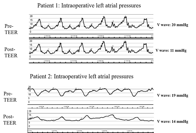
Figure 3.
Intraprocedural left atrial pressure wave tracings immediately before andafter transcatheter edge-to-edge mitral valve repair for both patients.The left atrial tracings were performed using the steerable guide catheterduring the MitraClip XTR procedure immediately before and after clipplacement under general anesthesia conditions. TEER: transcatheteredge-to-edge mitral valve repair