

Figure 1
Post-Ross right-ventricle-to-pulmonary-artery (RV-PA) homograft. (A) Ross procedure schematic. Source: Medical gallery of Blausen Medical, WikiJournal of Medicine, Wikiversity. (B) Cardiovascular magnetic resonance imaging demonstrated severe RV-PA homograft stenosis with high velocity jet directed toward the pulmonary artery (PA). LV: left ventricle

Figure 2
Balloon dilation of right-ventricle-to-pulmonary-artery (RV-PA) homograft. (A) Lateral view of left ventricular outflow tract (LVOT) angiogram during 24-mm balloon inflation. The left coronary artery (LCA) was not well seen (yellow arrow). (B) Neoaortic (Ao) root angiogram post-balloon deflation demonstrates contrast extravasation from the left coronary ostium to the pulmonary artery (PA). (C) Frontal view of LVOT angiogram in retrospect demonstrates compression of the neoaorta by the inflated balloon rather than poor contrast filling due to obstructed cardiac output. RCA: right coronary artery; LV: left ventricle

Figure 3
Prosthetic pulmonary valve stenosis following surgical replacement. On extracorporeal membrane oxygenation, the newly-placed surgical bioprosthetic pulmonary valve exhibited high velocity (5 m/s) on transesophageal echocardiogram: (A) two-dimensional, (B) color, and (C) continuous-wave Doppler.
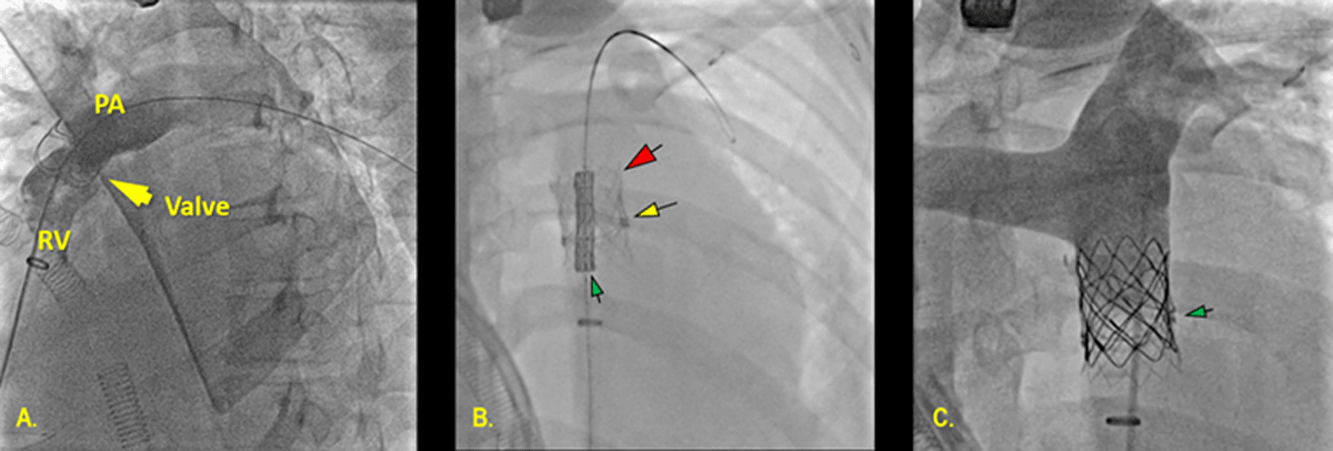
Figure 4
Hybrid pulmonary valve replacement. (A) Intraoperative right ventricular outflow tract angiogram demonstrated restrictive orifice and anterior filling defect. (B) A Palmaz P4010XL stent (red arrow) deployed across the prosthetic pulmonary valve (yellow arrows) completely ablated the orifice restriction. (C) Post-valve angiography demonstrated competency of the Melody valve (green arrows). PA: pulmonary artery; RV: right ventricle
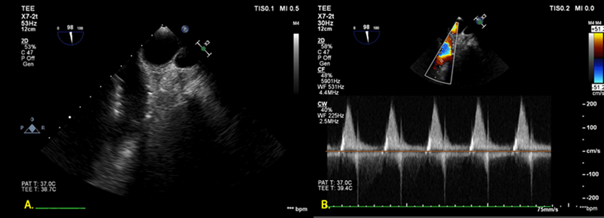
Figure 5
Postoperative echocardiogram. Post-hybrid valve transesophageal echocardiogram demonstrated normal velocity and pressure gradient across the pulmonary valve.