
Figure 1
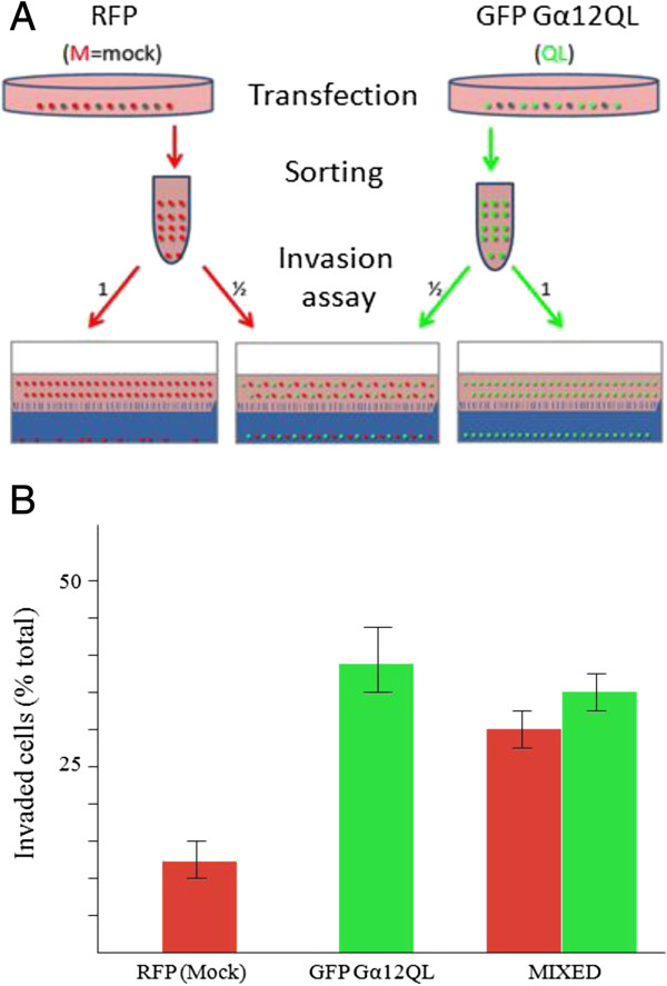
Dominant active Gα12 increases invasiveness of MDA-MB-231 breast cancer cells via paracrine signaling. (A) Experimental scheme of the in vitro invasion assay to test hypothesis that MDA-MB-231 cells transfected with Gα12QL drive the invasion of neighboring untransfected cells. Following transfection with the indicated vectors, cells were sorted and enriched fractions of RFP/mock (M) or GFP/Gα12QL (QL) containing cells (each at ~98% purity), or a 1:1 mixture of these cells, were subjected to the invasion assay followed by FACS analyses. (B) Results from FACS analysis showing invasion of the population of RFP/mock, GFP/Gα12QL and a mixed population of both RFP/mock and GFP/Gα12QL cells. Invaded cells were counted and plotted as a percentage of total cells subjected to the analysis. Values are plotted as the mean ± S.E. The results are from a single experiment that is representative of three independent experiments.
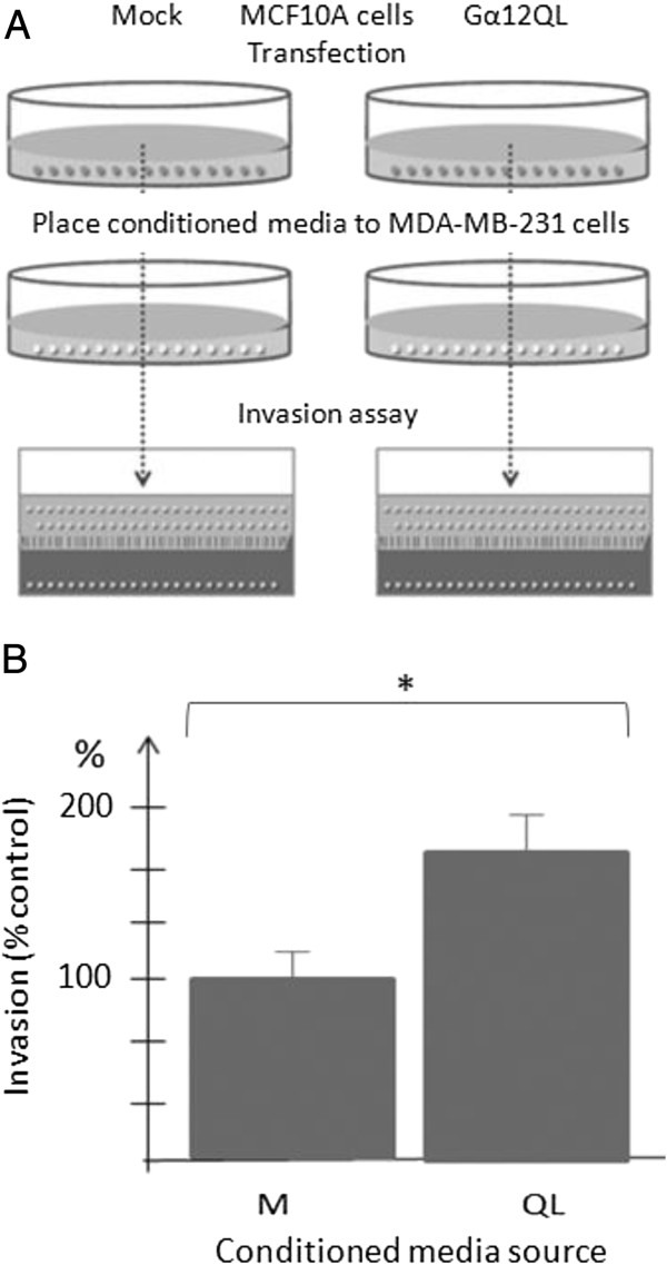
Figure 2
Factors secreted from MCF10A cells expressing dominant active Gα12 stimulate the invasion of MDA-MB-231 cells. (A) Experimental scheme illustrating experimental conditions. MCF10A cells were transfected as described under “Methods”. Following a 48 h incubation period, the conditioned media was collected and placed on MDA-MB-231 cells in 6 well plates. After 12 h, the MDA-MB-231 cells were harvested and subjected to an invasion assay. The results are shown in (B). Data are presented as a mean of triplicate determinations from a single experiment that is representative of two independent experiments. Bars represent the mean ± S.E. *p < 0.05.
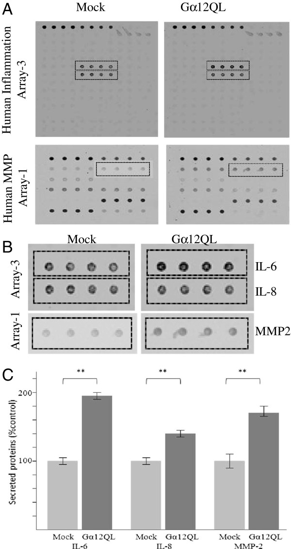
Figure 3
Expression of dominant active Gα12 in MDA-MB-231 cells induces secretion of cytokines IL-6 andIL-8, and MMP-2. (A) Protein array analysis of factors present in conditioned media. MDA-MB-231 cells were transfected either with control vector (Mock) or Gα12QL as indicated. Following a 72 h incubation period, media was harvested and subject to antibody-based arrays; see Methods for details. (B) Enlarged areas of the indicated regions of the arrays shown in (A). Quantification of data from three arrays is shown in (C). Bars represent the mean ± S.E. of quadruplicate determinations; IL-6 (p = 0.007), IL-8 (p = 0.003), MMP-2 (p = 0.007).
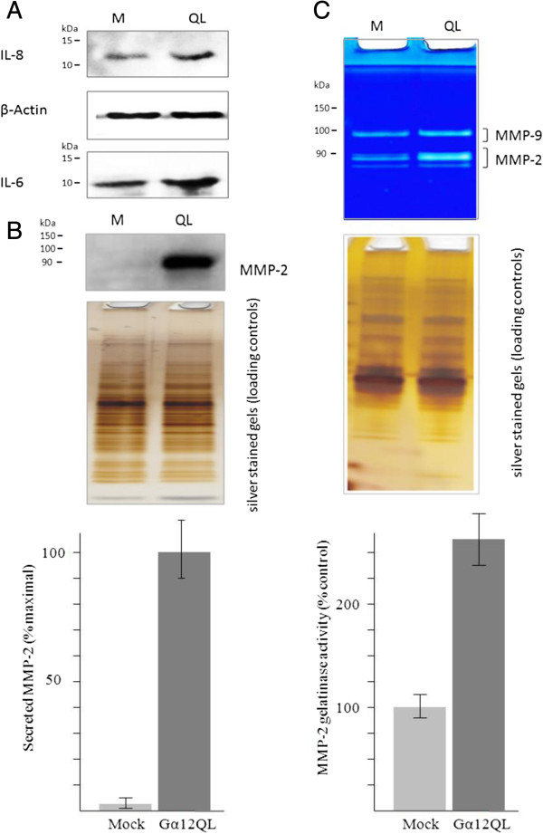
Figure 4
Validation of increased secretion of IL-6, IL-8 and MMP-2 expression of MDA-MB-231 cells expressing dominant activate Gα12. (A) Expression of IL-6 and IL-8 in total cell lysates of MDA-MB-231 cells transfected with either mock (M) vector or Gα12QL (QL) vector. MDA-MB-231 cells were transfected with the indicated construct and cell lysates prepared 72 h later and subject to immunoblot analysis as described under Methods. (B) MMP-2 levels in conditioned media of MDA-MB-231 cells transfected with either mock (M) vector or Gα12QL (QL) vector. MDA-MB-231 cells were transfected with the indicated construct and media harvested 72 h later and subject to immunoblot analysis as described under Methods. (C) Zymography analysis of gelatinase activity of MMP-2 in conditioned media of MDA-MB-231 cells transfected with either mock (M) vector or Gα12QL (QL) vector. Experimental conditions are as in (B). In the center panel, aliquots of conditioned media from were analyzed by silver-as the loading controls. Data in the lower panel are the mean ± S.E. of quadruplicate determinations from a single experiment that is representative of two independent experiments.
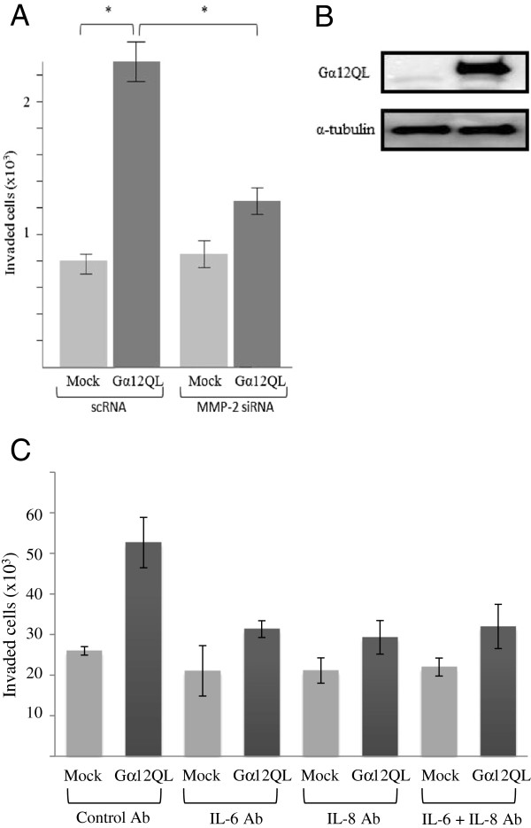
Figure 5
Interleukins and MMP-2 are involved in Gα12-mediated invasion of MDA-MB-231 cells. MDA-MB-231 cells were transfected with either control vector (Mock) or Gα12QL vectors as indicated. In addition, for (A) the cells were co-transfected with either scrambled (scRNA) or MMP-2 siRNA; the plasmid vectors also expressed GFP. The green cells were then sorted, and the enriched fractions were subjected to invasion assays. Depletion of MMP-2 with specific siRNA decreases Gα12-stimulated invasion of MDA-MB-231 cells. Data is pooled from three independent experiments, each involving duplicate determinations. Bars represent the mean ± S.E.; *(p < 0.05). (B) Representative western blot showing the over-expression of Gα12QL after doxycycline induction in MDA-MB-231 stable cell line. (C) Addition of IL-6 and IL-8 specific antibodies to the media of MDA-MB-231 cells stably expressing Gα12QL impairs Gα12-stimulated cell invasion. Data is pooled from three independent experiments, each involving triplicate determinations. Bars represent the mean ± S.E.
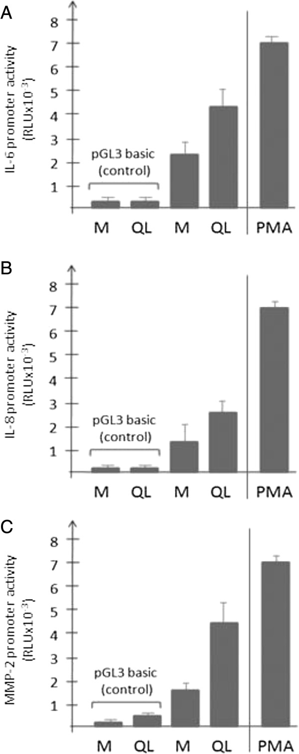
Figure 6
Ectopic expression of dominant active Gα12 in MDA-MB-231 cells enhances promoter activities of IL-6, IL-8 and MMP-2. Luciferase-based assays were conducted to analyze the impact of Gα12QL expression on the activities of an IL-6-Luc promoter (A), IL-8-Luc promoter construct (B), and a MMP-2-Luc promoter construct. (C) Mock (M) or Gα12QL expression vectors (200 ng of DNA per well of a 24-well plate) were co-transfected into the MDA-MB-231 cells along with the IL-6-Luc, IL-8-Luc or MMP-2-Luc vectors respectively, as well as pRL-TK (renilla control) as described in Methods. Induction of promoter activity by dominant active Gα12 expression or 10 ng/ml PMA (positive control) was assessed for the respective constructs, corrected for transfection efficiency, and calculated as percentage of induction compared with unstimulated baseline activity. Data shown were pooled from three independent experiments, each with triplicate determinations. Bars represent the mean ± S.E.
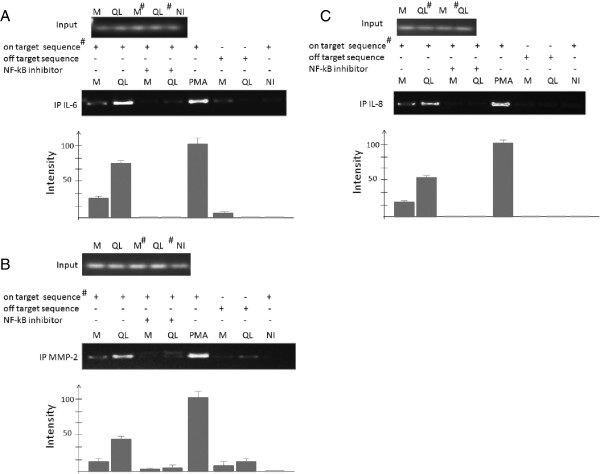
Figure 7
ChIP assays reveals Gα12QL-dependent increase of NF- κ B occupancy of IL-6, IL-8, and MMP-2 promoters. ChIP assays were performed on chromatin isolated from MDA-MB-231 cells transfected with mock (M) or Gα12QL vector expression vectors. Samples were pre-cleared by a precipitation protocol using control IgG, and then subjected to immunoprecipitation with anti-NF-κB. Semi-quantitative PCR was performed with primers targeted to a consensus NF-κB binding site upstream of the 5′-end of the corresponding genes. The sequences of neighboring regions were used as the “off target” sequence. The quantitative data reflect occupancy and represent mean ± SE of three independent experiments.