
Figure 1
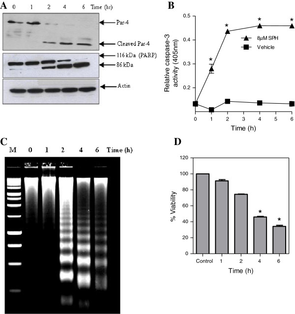
Sphingosine induces Par-4 cleavage in Jurkat cells. Panel A, Jurkat cells were treated with 8 μM SPH for the indicated time period and western blot analysis of cell lysate was carried out using an antibody against Par-4. Apoptosis induction was confirmed by western blot analysis of PARP cleavage using an antibody recognizing full-length and cleaved form of PARP. Control of protein loading was confirmed by using an antibody against actin. Panel B, Jurkat cells treated with SPH (8 μM) for the indicated time period and enzymatic activity of caspase-3 was measured as described in the materials and methods. Data shown are means ± SD (n = 3). *, p < 0.05 compared with untreated control. Panel C, Jurkat cells were treated with SPH (8 μM) for indicated time period and DNA fragmentation was analysed as described in the materials and methods. Panel D, WST cell viability assay for Jurkat cells following treatment with SPH (8 μM) for indicated time. Data shown are means ± SD (n = 3). *, p < 0.05 compared with untreated control.
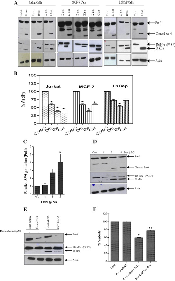
Figure 2
Par-4 cleavage is a common step in the process of caspase-dependant apoptosis. Panel A, Jurkat cells (0.2 μM Dox, 4 μM Eto and 25 μM Cur), MCF-7 cells (3 μM Dox, 100 μM Eto and 40 μM Cur) and LNCaP cells (5 μM Dox, 50 μM Eto and 50 μM Cur) were treated for 24 h and cleavage of Par-4 was analyzed by western blot analysis using Par-4 antibody. Apoptosis induction was confirmed by PARP cleavage and actin was used as protein loading control. Panel B, Jurkat, MCF-7 and LNCaP cells were treated with Dox, Eto and Cur for 24 h and cell viability was measured by using WST viability assay kit. Data shown are means ± SD (n = 3). *, p < 0.05 compared with untreated control. Panel C, MCF-7 cells were treated with indicated concentration of Dox and intracellular SPH generation was measured. Data shown are means ± SD (n = 3).*, p < 0.05 compared with untreated control. Panel D, MCF-7 cells were treated with indicated concentration of Dox, cells were lysed and Western blot analysis of Par-4 and PARP were carried out. Actin was used as a loading control. Panel E, MCF-7 cells were transiently transfected with control siRNA (Cont siRNA) and Par-4 siRNA. After 24 h, cells were treated with either vehicle or 3 μM Dox for further 24 h. The expression of Par-4 and PARP were detected by using Western blot analysis. Actin was used as the protein loading control. Panel F, MCF-7 cells were transiently transfected with Cont siRNA and Par-4 siRNA. After 24 h cells were treated with either vehicle or 3 μM Dox for further 24 h. The cell viability assay was performed. Data shown are means ± SD (n = 3). *, p < 0.05, and **, p < 0.05 compared with untreated control and control siRNA Dox treated cells, respectively.
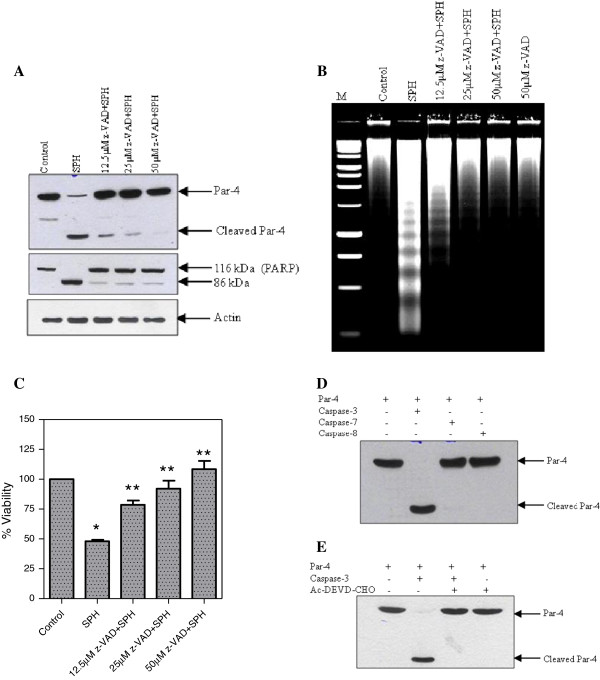
Figure 3
Cleavage of Par-4 by caspase-3 in vitro. Panel A, Effect of pan caspase inhibitor on the cleavage of Par-4. Jurkat cells were pre-treated with z-VAD-fmk at the indicated concentration for 1 h. The cells were then treated with SPH (8 μM) for 6 h and the lysate were immunoblotted with Par-4 and PARP antibodies. Actin was used as the loading control. Panel B, Jurkat cells were pre-treated with z-VAD-fmk at the indicated concentration for 1 h, then the cells were treated with SPH (8 μM) for 6 h and DNA fragmentation was analysed. Panel C, Jurkat cells were pre-treated with z-VAD-fmk at the indicated concentration for 1 h, then the cells were treated with SPH (8 μM) for 6 h and cell viability was measured by using WST viability assay kit. Data shown are means ± SD (n = 3). *, p < 0.05, and **, p < 0.05 compared with untreated control and SPH treated cells, respectively. Panel D, Immuno purified Par-4 was incubated with 300 ng of each purified caspase-3, -7, and −8 for 2 h. The reaction was stopped by adding SDS-PAGE sample buffer and proteolytic cleavage of Par-4 was detected by using anti-Par-4 antibody. Panel E, Immuno purified Par-4 was incubated with 300 ng of purified caspase-3 in the presence and absence of caspase-3 specific inhibitor (Ac-DEVD-CHO). The reaction was stopped by adding SDS-PAGE sample buffer and proteolytic cleavage product of Par-4 was detected by using anti-Par-4 antibody.
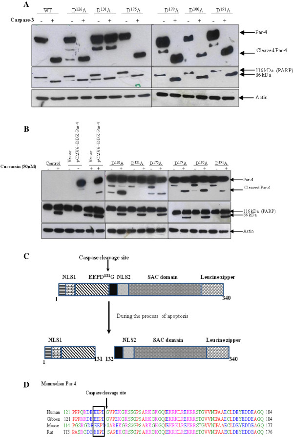
Figure 4
Identification of the cleavage site. Panel A, D126A, D131A, D175A, D179A, D180A, and D191A mutant were constructed. Each mutants were transiently transfected into HEK-293 cells. Cells were harvested 1 day after transfection and the cytosolic fraction of each transfectants were incubated with caspase-3 for 2 h in vitro. The proteolytic cleavage product of Par-4 was detected by anti-DDK monoclonal antibody. The activity of caspase-3 was also confirmed by checking the PARP cleavage. Actin was used as the loading control. Panel B, Wild type and each mutant Par-4 was transiently transfected in to HEK-293 cells and the cells were treated with either vehicle or 50 μM Cur for 24 h. The proteolytic cleavage of each mutant Par-4 was detected by immuno-probing of each lysate with anti-DDK monoclonal antibody. Apoptosis induction was confirmed by PARP cleavage and actin was used as protein loading control. Panel C, Schematic representation of the release of SAC domain containing fragment from the Par-4 during the apoptosis. Nuclear localization sequences (NLS1 and NLS2), selective for apoptosis induction in cancer cells (SAC). Panel D, Alignments of the regions containing the caspase-3 cleavage site of Par-4 from different species.
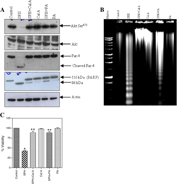
Figure 5
Inhibition of Akt dephosphorylation protects from SPH-induced Par-4 cleavage and apoptosis. Panel A, Jurkat cells were pre-treated with 5 nM calyculin A (Cal A) and 30 μM phosphatidic acid (PA) followed by treatment with 8 μM SPH. Cells were lysed and fractionated by SDS-PAGE. Western blot were probed with antibodies specific for phospho-Akt (Ser473), Akt, Par-4 and PARP. Actin was used as loading control. Panel B, Jurkat cells were pre-treated with 5 nM Cal A and 30 μM PA followed by treatment with 8 μM SPH and DNA fragmentation analysis was performed. Panel C, Jurkat cells were pre-treated with 5 nM CalA and 30 μM PA followed by treatment with 8 μM SPH and viability assay was done by using WST assay kit. Data shown are means ± SD (n = 3). *, p < 0.05, and **, p < 0.05 compared with untreated control and SPH treated cells, respectively.