
Figure 1
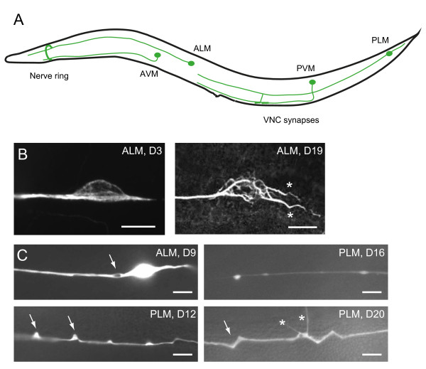
Age-dependent defects of C. elegans touch receptor neurons. (A) Schematic diagram of C. elegans touch receptor neurons, lateral view. For simplicity, only one of the bilateral ALM and PLM neurons was shown. VNC, ventral nerve cord. (B) Immunofluorescence of acetylated microtubules in the soma of young and old ALM neurons. Compared to the young neuron, the old ALM neuron showed aberrant sprouting from the soma (asterisks) and marked disorganization of microtubules. (C) Age-dependent axon defects in the touch neurons. ALM or PLM neurons were visualized in live animals with GFP expressed from the touch neuron-specific mec-4 promoter. Arrows mark bubble-like lesions (upper left, ALM), beading (upper right, PLM), blebbing (lower left, PLM) and wavy processes (lower right, PLM). Asterisks label neurite branching in the PLM.
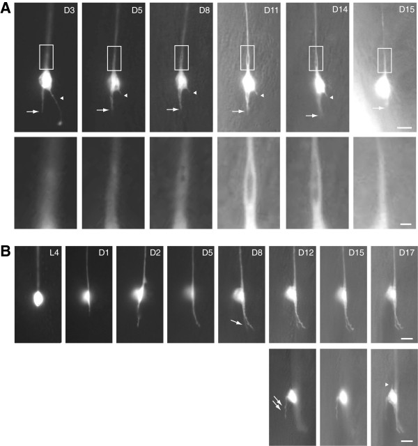
Figure 2
Temporal evolution of neuronal defects during aging in C. elegans. The same ALM neurons in the wild type were imaged at different time points over the animals’ lifespan; lateral view, anterior is up. Neurons were labeled by a touch cell-specific GFP reporter. Scale bar = 5 μm or 1 μm (A, insets). (A) The posterior process of the ALM neuron (arrow) remained static from D5 to D14 but retracted later. Arrowheads mark an ectopic sprouting from the soma, which was truncated between D3 and D5, and completely retracted on D15. Insets highlight the development of a bubble-like lesion in the proximal ALM process. The animal died on D16. (B) The ALM grew a posterior process on D1, which continued to lengthen between D1 and D5, and branched at D8 (arrow). An ectopic branch emerged from the dorsal side of the cell body at D12 (lower panel, double arrows). On D17, another short sprouting grew at the anterior aspect of the neuron (arrowhead). The three images of the right lower panel were taken from different focal planes. Images were originally published in the Proceedings of the National Academies of Sciences of the U.S.A. and reused with permission [30].
Table 1
C. elegans genes that regulate neuronal aging
| Genes | Identity | Major function |
|---|---|---|
| slo-1 | BK-type potassium channel | Muscle inactivation, neuronal excitability, etc |
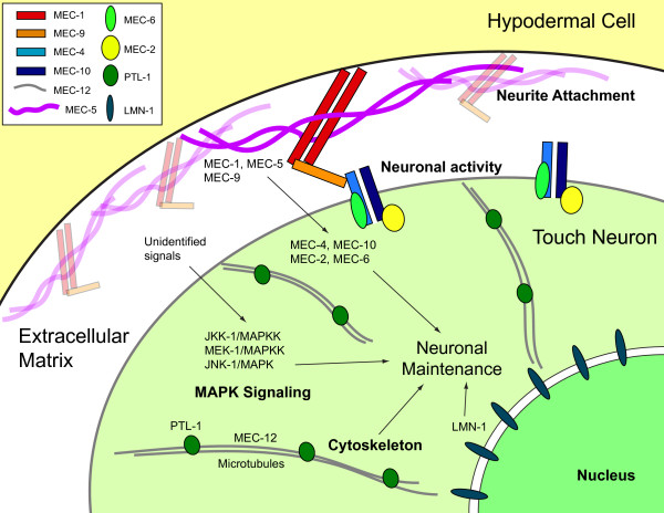
Figure 3
Schematic model of genetic and signaling networks that regulate maintenance and aging in C. elegans touch neurons. The touch neurons and their processes are ensheathed by the cytoplasmic extension of the neighboring hypodermal cell. Extracellular matrix containing the EGF- and Kunitz-domain proteins MEC-1 and MEC-9, and also atypical collagen MEC-5, was deposited between the touch neurons and the hypodermal cell. It is generally speculated that MEC-1, MEC-5 and MEC-9 tether the mechanosensory transduction channels, composed of MEC-2, MEC-4, MEC-6 and MEC-10, on the touch cell membrane and mechanically gate these channels. Although Tank et al. [31] had shown that components in the MAPK pathways, including JNK-1, JKK-1 and MEK-1, maintain touch neuron structures by inhibiting aberrant branching during aging, signals that activate these genes as well as their effectors or targets remain elusive. Genes that encode components of the microtubule cytoskeleton (MEC-12/α-tubulin and PTL-1/Tau) or the integral nuclear envelope protein (LMN-1/lamin) are also important for maintaining postmitotic neurons in C. elegans. For simplicity, this schematic diagram was generated in the form of the neuronal soma, but similar models could also apply to the process of the neuron.