
Figure 1
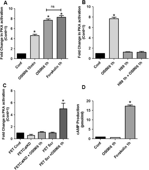
OSI-906 mediates cAMP independent and AKAP dependent PKA activation in colon cancer cells. FET cells treated with OSI-906 (1 μM) activated PKA in a time dependent fashion (A). Forskolin (10 μM) was used as a positive control. PKA inhibitor H89 (15 μM) was used to inhibit PKA activation. Pretreatment with H89 followed by OSI-906 treatment abrogated the PKA activation (B). Knockdown of PKA Catalytic subunit (FETCatKD) abrogated OSI-906 mediated PKA activation. However, FET cells transfected with Scrambled siRNA (FET Scr) cells showed PKA activation following OSI-906 treatment. (C). PKA activation by OSI-906 is cAMP independent as determined via cAMP assay (D). All experiments were repeated three times independently with 3 replicas per repeat for the ANOVA analysis. (* P < 0.001).
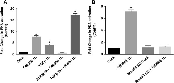
Figure 2
OSI-906 activates PKA in a TGFβ signaling dependent manner. FET cells pretreated treated with TGFβRI inhibitor ALK5i (400 nM) abrogated OSI-906 (1 μM) mediated PKA activation. FET cells treated with TGFβ (5 ng/ml) and OSI-906 together showed an increase in PKA activation (A). OSI-906 mediated PKA activation is Smad3-dependent as determined by knockdown of Smad3 in FET cells (B). All experiments were repeated three times independently with 3 replicas per repeat for the ANOVA analysis (* P < 0.001).
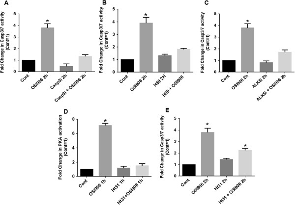
Figure 3
OSI-906 mediates apoptosis by a TGFβ/PKA dependent manner. FET cells treated with OSI-906 (1 μM) activated Caspase 3/7. Caspase 3 inhibitor C3i (20 μM) was used to inhibit Caspase 3 activation (A). Caspase 3/7 activation by OSI-906 is PKA dependent as activation of Caspase 3/7 was abrogated with prior treatment of FET cells with PKA inhibitor H89 (15 μM) (B). FET cells treated with TGFβRI inhibitor ALK5i (400 nM) showed abrogation of Caspase 3/7 activation. Pretreatment with ALK5i followed by OSI-906 showed similar results (C). Activation of PKA by OSI-906 was abrogated in the presence of 25 μM AKAP inhibitor Ht31 (D). Caspase 3/7 activation by OSI-906 is AKAP dependent as OSI-906 mediated Caspase 3/7 activation was abrogated with prior treatment of FET cells with Ht31 (25 μM) (E). All experiments were repeated three times independently with 3 replicas per repeat for the ANOVA analysis (* P < 0.001).
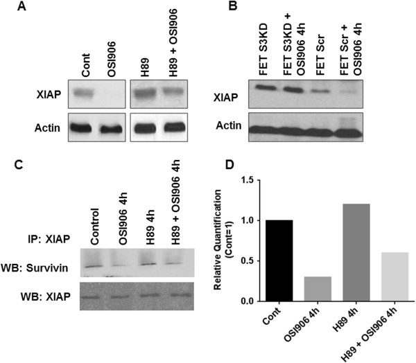
Figure 4
OSI-906 regulates XIAP in a TGFβ/PKA dependent manner. FET cells treated with OSI-906 (1 μM) showed effective downregulation of XIAP. This observed downregulation is PKA dependent as downregulation of XIAP was abrogated in FET cells treated with PKA inhibitor H89 (15 μM) (A). OSI-906 mediated XIAP downregulation is dependent on Smad3 as determined by shRNA knockdown of Smad3 in FET cells (termed as FETS3KD). FETS3KD cells treated with OSI-906 showed no change in XIAP expression. However, FET Scr cells showed XIAP downregulation following treatment with OSI-906 (B). FET cells treated with OSI-906 effectively dissociated XIAP from Survivin as determined by immunoprecipitation (IP) assay (C). OSI-906 mediated XIAP/Survivin dissociation is PKA dependent as dissociation was abrogated when FET cells were treated with H89 and/or pretreated with H89 prior to OSI-906 treatment. Relative quantification of the IP has been performed showing reduced interaction between survivin/XIAP following OSI-906 treatments (D).
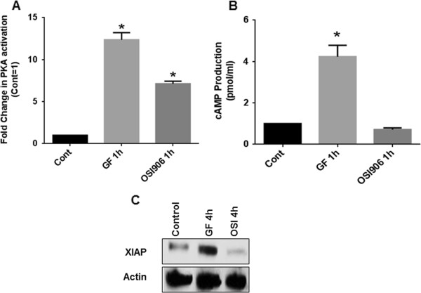
Figure 5
Differential PKA activation decides cell survival or apoptotic fate. FET cells treated with Growth Factors (or GF that includes a combination of Transferrin and Insulin) or OSI-906 (1 μM) activated PKA (A). FET cells treated with GF showed cAMP-dependent activation of PKA whereas FET cells treated with OSI-906 showed cAMP-independent activation of PKA as determined by cAMP assay (B). FET cells treated with GF showed an upregulation of XIAP, whereas cells treated with OSI-906 showed a downregulation of XIAP (C). All experiments were repeated three times independently with 3 replicas per repeat for the ANOVA analysis (* P < 0.001).
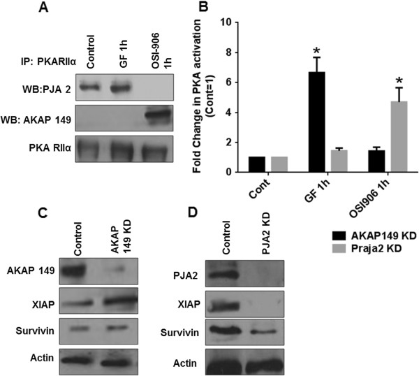
Figure 6
Different AKAPs compete to bind PKA based on cAMP-dependent or independent mode of PKA activation. FET cells treated with GF showed an effective increase in the association of Praja2 and RII subunit of PKA (PKARIIα) and a decrease in PKARII/Praja2 association when FET cells were treated with OSI-906. FET cells treated with OSI-906 showed an effective increase of AKAP149and PKARIIα association whereas an effective decrease in this association was observed when cells were treated with GF (A). OSI-906-mediated PKA activation requires AKAP149association with PKA as determined by knockdown of AKAP149in FET cells. PKA activation by GF treatment requires Praja2 association with PKA as determined by knockdown of Praja2 in FET cells (B). AKAP149is associated with negative regulation of IAPs survivin and XIAP as determined by knockdown of AKAP149in FET cells (C). Praja2 is associated with positive regulation of survivin and XIAP as determined by knockdown of Praja2 in FET cells (D). All experiments were repeated three times independently with 3 replicas per repeat for the ANOVA analysis (* P < 0.001).
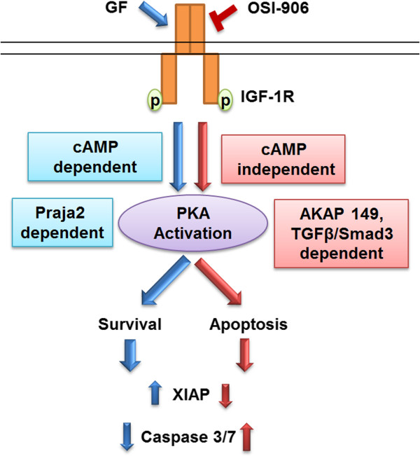
Figure 7
Schematic representation of AKAP/PKA signaling that regulates cell fate.