
Figure 1
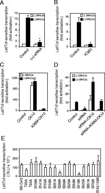
CK1δ positively regulates Wnt/β-catenin signaling. Panel A, siRNA targeting CK1δ attenuates Lef/Tcf-sensitive transcription. F9 cells were transfected with siRNA targeting CK1δ one day before co-transfection with Rfz1 and Super8xp TOPFlash (M50) reporter. On the following day, cells were either unstimulated or stimulated with Wnt3a for 7 hr. Cell lysates were assayed for Lef/Tcf-sensitive transcription. Results are displayed relative to the unstimulated cells (set to “1”). Results shown are mean values ± s.e.m. from 5 independent experiments. Statistical significance is indicated (*, p < 0.005). Panel B, treatment of CK1δ ιnhibitor attenuates Lef/Tcf-sensitive transcription. F9 cells were co-transfected with Rfz1 and M50. One day post transfection, cells were either untreated or pretreated with IC261 (20 μM, selective inhibitor of CK1δ/ϵ) for 1 hr prior to stimulation either with or without Wnt3a (20 ng/ml) for 7 hr in the continued presence (or absence) of IC261. Cell lysates were prepared and assay of Lef/Tcf-sensitive transcription was performed. Results are displayed relative to the unstimulated cells (set to “1”). Results shown are mean values ± s.e.m. from 3 independent experiments. Statistical significance is indicated (*, p < 0.005). Panel C, expression of CK1δ enhances Lef/Tcf-sensitive transcription. F9 cells were transfected with Rfz1, M50 and either wild-type CK1δ or K38M-CK1δ (kinase inactive mutant). On the following day, cells were either unstimulated or stimulated with Wnt3a for 7 hr. Cell lysates were assayed for Lef/Tcf-sensitive transcription. Results are displayed relative to the unstimulated cells (set to “1”). Statistical significance is provided, * denotes p < 0.005 (versus control cells). Results shown are mean values ± s.e.m. from 4 independent experiments. Panel D, wild-type CK1δ rescues Lef/Tcf-sensitive transcription in the CK1δ-deficient cells. F9 cells stably expressing Rfz1 and M50 were treated with siRNA targeting CK1δ one day before transfection with either wild-type CK1δ or K38M-CK1δ. On the following day, cells were either unstimulated or stimulated with Wnt3a for 7 hr. Cell lysates were assayed for Lef/Tcf-sensitive transcription. Results are displayed relative to the unstimulated cells (set to “1”). Statistical significance is provided, * denotes p < 0.005 (versus control cells). Results shown are mean values ± s.e.m. from 3 independent experiments. Panel E, role of serine/threonine sites of Dvl2 in Lef/Tcf-sensitive transcription. Phosphorylation of rDvl2 (purified from Sf9 cells) catalyzed by purified CK1δ was carried out in vitro and phosphorylation sites were identified by mass spectrometry. The phosphorylation sites (S/T) identified then were mutated to alanine using Quickchange Mutagenesis System (Stratagene, La Jolla, CA). F9 cell were co-transfected with Rfz1, M50 and either wild-type Dvl2 or a specific Dvl2 mutants. On the following day, assay for Lef/Tcf-sensitive transcription was performed. Statistical significance is provided, * denotes p < 0.005 (versus wild-type Dvl2 expressed cells). Results shown are mean values ± s.e.m. from 6 independent experiments.
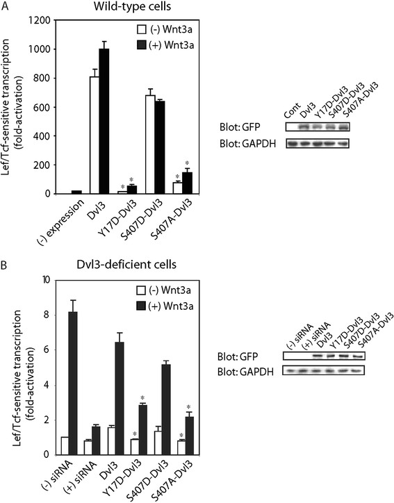
Figure 2
Interrogation of the functional status of Y17 and S407 sites of Dvl3 in Lef/Tcf-sensitive transcriptional activation in response to stimulation with Wnt3a. Panel A, F9 cells co-expressed with Rfz1, Super8xTOPFlash (M50) and either wild-type Dvl3 or one of following Dvl3 mutants (Y17D-Dvl3, S407A-Dvl3 and S407D-Dvl3) were stimulated with or without Wnt3a for 7 hr. Cell lysates were assayed for Lef/Tcf-sensitive transcription. Cell lysates were immunoblotted and subsequently stained with anti-GFP (Dvl3 expression) and anti-GAPDH (control) antibody. Results are displayed for transcriptional activity relative to unstimulated cells (set to “1”). Statistical significance is indicated (*, p < 0.005, versus Dvl3 expressed cells). Panel B, F9 cells stably expressing Rfz1 and M50 were treated with siRNA targeting Dvl3 one day before transfection of the cells with either wild-type Dvl3 or Y17D-Dvl3 or S407A-Dvl3 or S407D-Dvl3. On the following day, cells were stimulated with Wnt3a for 7 hr. Cell lysates were assayed for Lef/Tcf-sensitive transcription. Cell lysates were immunoblotted with either anti-GFP or anti-GAPDH antibody (control). Results are displayed for transcriptional activity relative to unstimulated cells (set to “1”). Statistical significance is indicated (*, p < 0.05, versus Dvl3 expressed cells).
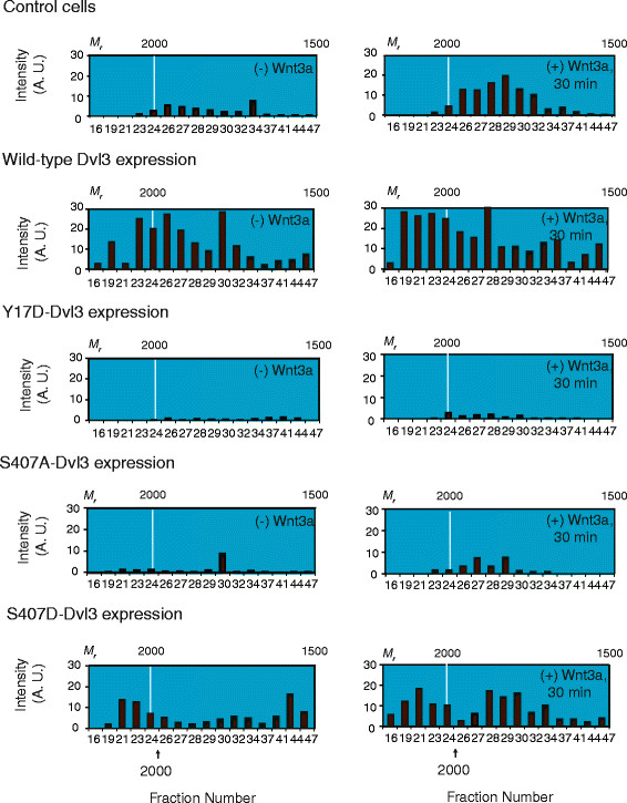
Figure 3
Interrogation of functional roles of Y17 and S407 sites of Dvl3 in assembly of very large Dvl3-based supermolecular complexes in response to stimulation with Wnt3a. Y17D-Dvl3 and S407A-Dvl3 abolished the assembly of Dvl3-based supermolecular complexes in response to Wnt3, whereas S407D-Dvl3 enhances the assembly of Dvl3-based supermolecular complexes. Cells expressing either wild-type Dvl3 or Y17D-Dvl3 or S407A-Dvl3 or S407D-Dvl3 were stimulated with or without Wnt3a for 30 min. Cells were lysed and extracts (20 mg protein) were applied to Sephacryl S-400 gel filtration column (AKTA, GE Health Care). Fractions were analyzed by SDS-PAGE and immunoblotted, subsequently stained with anti-Dvl3 antibody. Dvl3 blots were quantified by the calibrated scanner and results were displayed. The Dvl3-based supermolecular complexes in F9 cells expressing Rfz1 were also displayed as a control. The calculated, relative molecular weight (Mr) positions from the calibration curve are labeled at the top. The bottom numbers indicate fraction number. Arrow indicates the precise position at which calibration proteins elute from Sephacryl S-400. Results are representative of at least 2 independent experiments.
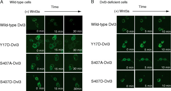
Figure 4
Interrogation of functional roles of Y17 and S407 sites of Dvl3 in formation of macromolecular Dvl3-based punctae in response to stimulation with Wnt3a. Panel A, live cell images of expression of either wild-type Dvl3 or Y17D-Dvl3 or S407A-Dvl3 or S407D-Dvl3 in response to Wnt3a. F9 cells were co-transfected with Rfz1 and either wild-type Dvl3 or Y17D-Dvl3 or S407A-Dvl3 or S407D-Dvl3. One day post transfection, cells were stimulated with Wnt3a and cells images were taken at 0, 15, and 30 min post stimulation with Wnt3a. Panel B, live cell images of cells expressing either wild-type or Dvl3 mutants (Y17D-Dvl3 or S407D-Dvl3 or S407D-Dvl3) in Dvl3-deficient cell: response to Wnt3a. F9 cells stably expressing with M50 and Rfz1 were treated with siRNA targeting Dvl3. On the following day, cells were transfected with either wild-type Dvl3 or one of the Dvl3 mutants. Post 24 hr of transfection, cells were stimulated with Wnt3a and live cell images were captured during Wnt stimulation. Results were displayed at 0, 5, and 10 min time points. The images shown are representative of three or more separate experiments.
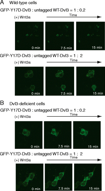
Figure 5
Formation of Dvl3-based punctae by wild-type versus mutant forms of Dvl3. Y17D-Dvl3 forms punctae in cells co-expressing wild-type Dvl3 in untreated and Dvl3-deficient F9 cells. Panel A, F9 cells were co-transfected with GFP- and HA-tagged Y17D-Dvl3 and increasing amount of co-expressed untagged wild-type Dvl3 (ratio = 1 : 0.2, or 1 : 2). Post 1 day transfection, cells were stimulated with Wnt3a. Cell images were taken during Wnt stimulation. Results were displayed at 0, 7.5, and 15 min time-points. Panel B, parallel experiments were performed in F9 cells made deficient of endogenous wild-type Dvl3 by siRNA. The images shown are representative of three or more separate experiments.
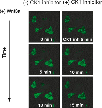
Figure 6
CK1δ/ϵ regulates formation of Dvl3-based very large punctae. Treatment with CK1δ/ϵ inhibitor decreases number as well as apparent size of the punctae. F9 cells were co-transfected with Rfz1 and GFP- and HA-tagged wild-type Dvl3. Cells were stimulated with Wnt3a and images of live cells were captured at the times indicated. After 10 min post Wnt stimulation, a CK1δ/ϵ selective inhibitor (IC261, 5 μM) was added and cell images captured continuously. Results displayed are images captured at 5, 10, and 15 min post treatment with IC261. The images displayed are representative of two or more separate experiments.
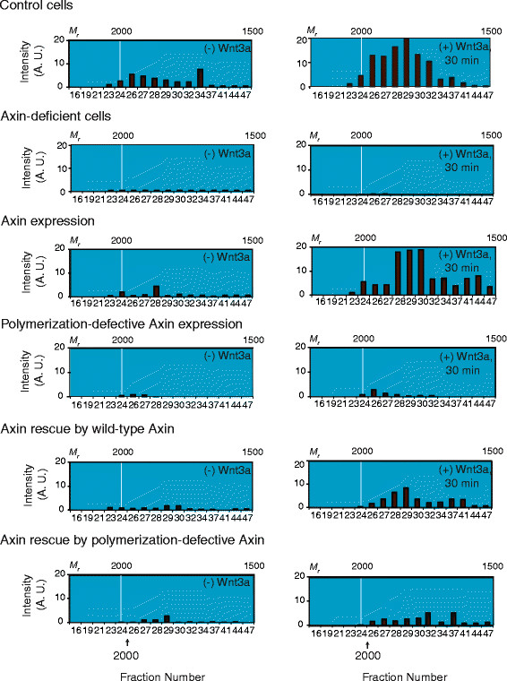
Figure 7
Expression of Axin M3 mutant precludes assembly of very large Dvl3-based supermolecular complexes in response to Wnt3a. Control cells, Wnt3a stimulates formation of very large Mr-complexes. SEC chromatogram of Dvl3-based complexes prepared from untreated cells. Axin-deficient cells, knockdown of Axin attenuates assembly of Dvl3-based supermolecular complexes in response to Wnt3a. F9 cells were transfected with siRNA targeting Axin one day before transfection with Rfz1. Twenty four hr later, cells were either unstimulated or stimulated with Wnt3a for 30 min. Axin expression, expression of wild-type Axin alone stimulates assembly of Dvl3-based supermolecular complexes. F9 cells were co-transfected with Rfz1 and expression vectors harboring human wild-type Axin. Two days post transfection, F9 cells were treated either without or with Wnt3a for 30 min. Polymerization-defective Axin expression, expression of M3 Axin mutant blocks assembly of very large Dvl3-based supermolecular complexes in response to Wnt3a. F9 cells were co-transfected with Rfz1 and expression vectors harboring human M3 Axin mutant. Two days post transfection, F9 cells were treated either without or with Wnt3a for 30 min. Rescue of Axin-depletion by expression of wild-type Axin, expression of wild-type Axin rescues the inability of Axin-deficient cells to assemble Dvl3-based supermolecular complexes in response to Wnt3a. Cells were treated with siRNA targeting Axin one day before subsequent co-transfection with Rfz1 and human wild-type Axin for an additional day. Twenty four hr after this final transfection, cells were either unstimulated or stimulated with Wnt3a for 30 min. Axin rescue by polymerization-defective Axin, expression of M3 Axin fails to rescue the inability of Axin-deficient cells to assembly of Dvl3-based supermolecular complexes in response to Wnt3a. Cells were treated with siRNA targeting Axin one day before subsequent co-transfection with Rfz1 and M3 Axin mutant for an additional day. Twenty four hr after this final transfection, cells were either unstimulated or stimulated with Wnt3a for 30 min. For all experiments, cell lysates were subjected to Sephacryl S-400 gel filtration column chromatography (SEC) and resolved fractions were analyzed by SDS-PAGE, immunoblotting and eventual staining with anti-Dvl3 antibody. Dvl3 blots were subjected to quantification by calibrated scanning and the results displayed are representative of at least 2 independent experiments, which yielded quantitatively similar results. The calculated, relative molecular weight (Mr) positions from the calibration curve are labeled at the top of each SEC chromatogram. The bottom labels indicate fraction number. Arrows indicate the precise position at which calibration protein elute from Sephacryl S-400.