
Table 1
BCL-2 family proteins
| BH3 only pro-apoptotic proteins | Multi-domain pro-apoptotic | Anti-apoptotic proteins |
|---|---|---|
| BNIP3 |
Of the BH3 only proteins, BID, BIM and possibly PUMA are considered apoptosis activators and the others as apoptosis sensitizers. BOK is an apoptosis effector expressed primarily in the ovary.
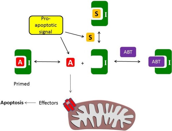
Figure 1
The intrinsic apoptosis pathway. Apoptosis is triggered by the formation of a pore in the mitochondrial outer membrane formed from the effectors. This is controlled by the activator molecules (BID, BIM, PUMA, labelled “A”) which directly stimulate the effectors (BAK and BAX). A pro-apoptotic signal (such as oncogene stress or chemotherapy) induces the expression of apoptosis activators and sensitizers. The activators may be sequestered by the inhibitors (labelled “I”, e.g. BCL-2, BCL-XL, MCL-1) and this prevents apoptosis. However, sensitizer molecules (labelled “S”, eg NOXA, BAD) or BH3 mimetics (eg ABT-737, labelled “ABT”) can occupy the inhibitors, preventing them from binding the activators. In cells in which the inhibitors are already primed with an activator, the effective displacement of the activator from the inhibitor by ABT-737 induces apoptosis.
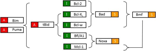
Figure 2
Binding specificity of BH3 only proteins. The activators (denoted with an “A”), BIM and PUMA bind to all the apoptosis inhibitors (denoted with an “I”). However, tBID and the sensitizers (denoted with a “S”) bind to a subset of the apoptosis inhibitors. There are some discrepancies in the literature (e.g., whether BMF binds MCL-1) and it is important to note that this diagram summarizes interactions (adapted from [7-9] in a binary fashion (interaction or no interaction) whereas in reality the interactions are better described by a range of binding affinities.
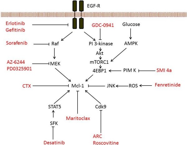
Figure 3
Strategies to decrease the expression of MCL-1. The sensitivity of cells to ABT-737 can be increased using drugs which decrease the expression of MCL-1. The ERK (including RAF and MEK) and PI 3-kinase pathways are key regulators of MCL-1 expression so drugs inhibiting this pathway show synergy with ABT-737. Drugs inhibiting other regulators of MCL-1 are also shown, and these drugs also increase sensitivity to ABT-737. ARC, 4-amino-6-hydrazino-7-beta-D-ribofuranosyl-7 H-pyrrolo(2,3-d)-pyrimidine-5-carboxamide; CTX, chemotherapy; EGF-R, EGF receptor; ROS, reactive oxygen species; SFK, SRC family kinase. For references, the reader is referred to the main text.