
Figure 1
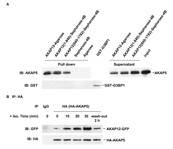
Docking of AKAP5 to full-length AKAP12 as well as to N-(1-840) and C-(840-1782) terminal fragments. A, Purified HA-tagged AKAP12 was immobilized to agarose beads, His-tagged AKAP12 (1-840) or His-tagged AKAP12 (840-1782) were cross-linked individually to CNBr-activated Sepharose 4B beads. Each of these bead types was incubated with either purified His-tagged AKAP5 or an equivalent amount of purified GST-tagged G3BP1 protein, as a control. At the end of the incubation the beads were collected from the mixtures and washed. The AKAP5 released from binding to sequences immobilized to the beads (Pull down), the AKAP5 input to the incubation (Input), and the AKAP5-depleted supernatants of the incubation post affinity adsorption (Supernatant) were sampled and applied to SDS-PAGE, subjected to immunoblotting and stained with anti-AKAP5 (upper panel) or anti-GST (lower panel) antibodies. B, A431 cells stably expressing GFP-tagged AKAP12 were transiently transfected with HA-tagged AKAP5. Confluent cells were treated with 10 μM isoproterenol for 10, 20, 30 min or treated for 30 min with isoproterenol and then washed-free of agonist for 2 h (wash-out 2 h). Whole-cell lysates were prepared and pull-downs conducted with anti-HA antibody (targeting HA-tagged AKAP5) or control IgG. The immune complexes were subjected to SDS-PAGE, transferred to PVDF membrane and probed with antibodies against HA tag (HA-AKAP5) or GFP (AKAP12-GFP). The results shown are taken from a single experiment, representative of at least three separate experiments performed on separate cultures of cells.
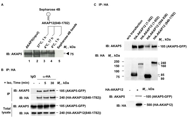
Figure 2
Docking of AKAP5 to AKAP12. A, Purified His-tagged AKAP12 (840-1782) was cross-linked to CNBr-activated Sepharose 4B, then incubated with purified His-tagged AKAP5 under the conditions labeled on each lane: lane 1, purified AKAP5, input; lane 2, pull-down at 37°C, 1 h; lane 3, pull-down at 4°C, 17 h; lane 4, pull-down at 4°C, 1 h; and lane 5, pull-down control, Sepharose 4B beads alone. B, A431 clones stably expressing GFP-tagged AKAP5 were transiently transfected to express HA-tagged AKAP12 C-(840-1782) terminal fragment. Confluent cells were treated with 10 μM isoproterenol for the indicated times. Whole-cell lysates were prepared and pull-downs performed with antibodies against the HA tag which were covalently linked to protein A/G-agarose beads or control IgG. The immune complexes were subjected to SDS-PAGE, transferred to PVDF membrane, and probed with antibodies against either AKAP5 or the HA tag. C, A431 cells stably expressing GFP-tagged AKAP5 were transiently transfected with HA-tagged AKAP12 or the AKAP12 fragments listed (i.e., 1-362; 1-652; 654-938; and 840-1782). The whole-cell lysates were prepared and pull-downs performed with antibodies against the HA tag which were covalently linked to protein A/G-agarose beads. The immune complexes were subjected to SDS-PAGE, transferred to PVDF membrane, and probed with antibodies against either AKAP5 or the HA tag. The results shown are taken from a single experiment, representative of at least three separate experiments performed on separate cultures of cells.
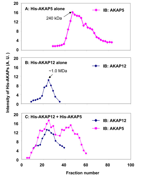
Figure 3
Steric-exclusion chromatography of AKAP5 and AKAP12 alone and in combination: identification of supermolecular AKAP5/AKAP12 hetero-oligomers. To ascertain the possible presence of higher-order oligomers of AKAP12 and AKAP5, steric-exclusion chromatography of purified AKAP5 and AKAP12 individually (A and B, respectively) or after incubation in combination (C) was performed on an AKTA FPLC (GE Healthcare) fitted with a HiPrep Sephacryl S-400 High resolution 16/60 column (GE healthcare). Marker proteins mobilities were employed to establish the MW of AKAP5-, AKAP12-, and AKAP5/AKAP12 supermolecular complexes by elution position. The A280 absorbance was monitored in real time. Samples from each fraction were subjected to SDS-PAGE and immunoblotting and staining for the AKAP12 or AKAP5. The results displayed are representative of at least three separate experiments performed on as many individual protein preparations.
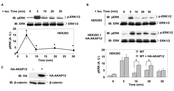
Figure 4
Expression of AKAP12 potentiates Erk1/2 activation by beta-adrenergic agonist in HEK293 cells. The cells were transiently transfected with HA-AKAP12 for 42 h and then changed to serum-free medium for 4 h. A, time course of ERK phosphorylation in HEK293 cells stimulated with 10 μM isoproterenol. B, overexpression of HA-AKAP12 potentiates ERK activation in HEK293 cells stimulated in response to 10 μM isoproterenol. C, Immunoblot analysis of expression of HA-AKAP12 using β-catenin as loading index. The blots shown are taken from a single experiment, representative of at least three separate experiments performed on separate cultures of cells. Tabular data from separate experiments was analyzed for variance using the Student's "t" test for significance. * p < 0.05.
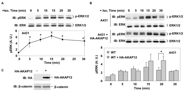
Figure 5
Expression of AKAP12 potentiates activation of Erk1/2 by beta-adrenergic agonist in A431 cells. A, time course of ERK phosphorylation in A431 cells stimulated with 10 μM isoproterenol. B, overexpression of HA-AKAP12 potentiates ERK activation in A431 cells stimulated with 10 μM isoproterenol. C, immunoblot analysis of expression of HA-AKAP12. The blots shown are taken from a single experiment, representative of at least three separate experiments performed on separate cultures of cells. Tabular data from separate experiments was analyzed for variance using the Student's "t" test for significance. * p < 0.05.
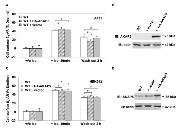
Figure 6
Effects of overexpression of AKAP5 on AKAP12-mediated recycling of desensitized, internalized β2-adrenergic receptors. HEK293 cells stably expressing β2-AR-GFP or wild-type A431 cells were transiently transfected with an expression vector harboring HA-AKAP5 or the empty vector for 24 h. The cells were untreated or treated with 10 μM isoproterenol for 30 min and then washed free of isoproterenol and allowed to recover for 2 h. Cell surface β2-AR content was detected with the cell impermeant radiolabeled beta-adrenergic antagonist [3H]-CGP12177. A, β2-AR recycling in A431 cells: effects of AKAP5 expression. B, expression of HA-AKAP5 in A431 cells analyzed by immunoblotting and staining with anti-AKAP5 antibody. C, β2-AR recycling in HEK293 cells: effects of AKAP5 expression. D, expression of HA-AKAP5 in HEK293 cells analyzed by immunoblotting and staining with anti-AKAP5 antibody. The results displayed are mean values ± SD derived of triplicates. For panels B and D, the results shown are taken from a single experiment, representative of at least three separate experiments performed on separate cultures of cells. Tabular data from separate experiments was analyzed for variance using the Student's "t" test for significance. # denotes p values > 0.05, i.e. differences that are not statistically significant.
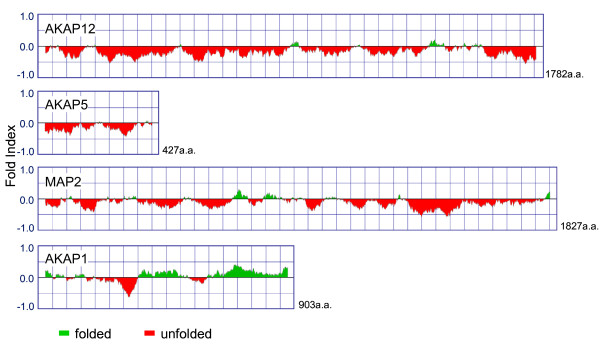
Figure 7
FOLDIndex© analysis of AKAP family members AKAP12, AKAP5, MAP2, and AKAP1: degree of natively "unordered" structure. The primary sequences of human AKAP5, AKAP12, and several other members of the AKAP family were analyzed for natively ordered structure by application of the FOLDindex©. The sequences are displayed from N-terminus to C-terminus (left to right) for four of the AKAP sequences selected. The regions below the horizontal midline, filled in with red, are predicted to be natively unfolded based upon primary sequence information alone. The regions above the horizontal midline, filled in with green, are predicted to be natively folded, based upon primary sequence.