
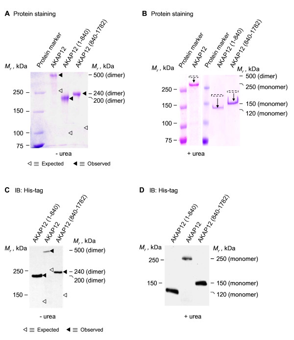
Figure 1
Oligomerization of AKAP12 (and either C-terminal or N-terminal fragments) expressed and purified from E. coli. His-tagged AKAP12 or His-tagged AKAP12 fragments were expressed in E. coli. The AKAP12 (or fragments) were purified and then resolved on SDS-PAGE in the absence (A) or presence of 8 M urea (B). The gels were fixed and stained for protein with Coomassie Brilliant Blue (A, B) or the resolved proteins transferred to PVDF membrane and subjected to immunoblotting (C, D). The "◀" indicates the observed Mr on the gels and the "Δ" indicates the predicted Mr, calculated from the primary sequence.
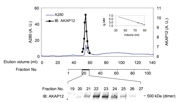
Figure 2
Steric-exclusion chromatography of purified AKAP12. His-tagged AKAP12 was purified and then resolved by steric-exclusion chromatography performed on an AKTA FPLC fitted with HiPrep Sephacryl-S400 (16/60) column. The presence of supermolecular oligomers of AKAP12 was established by SDS-PAGE and immunoblotting of samples from the chromatography. The resolved, transferred proteins were stained with anti-AKAP12 antibodies. Marker protein mobilities were employed to establish the Mr of AKAP12 by elution position (inset). The results displayed are representative of three separate experiments performed on as many separate cell cultures.
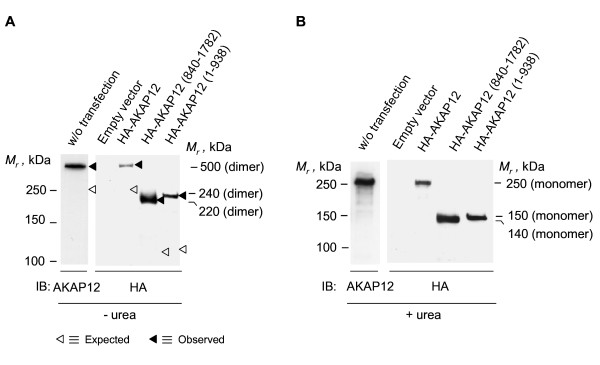
Figure 3
Oligomerization of AKAP12 (and either C-terminal or N-terminal fragments) expressed in mammalian HEK293 cells. Human embryonic kidney (HEK) 293 cells were transfected with an empty expression vector or one harboring either full-length AKAP12, AKAP12 N-terminal (1-938) or C-terminal (840-1782) regions. Whole-cell lysates of cells expressing either AKAP12 or one of the fragments were subjected to SDS-PAGE in the absence (A) or presence of 8 M urea (B). The resolved proteins were transferred to PVDF membrane and subjected to immunoblotting, stained for AKAP12 or the HA-tag. The results displayed are representative of at least three separate experiments performed on as many separate cell cultures.
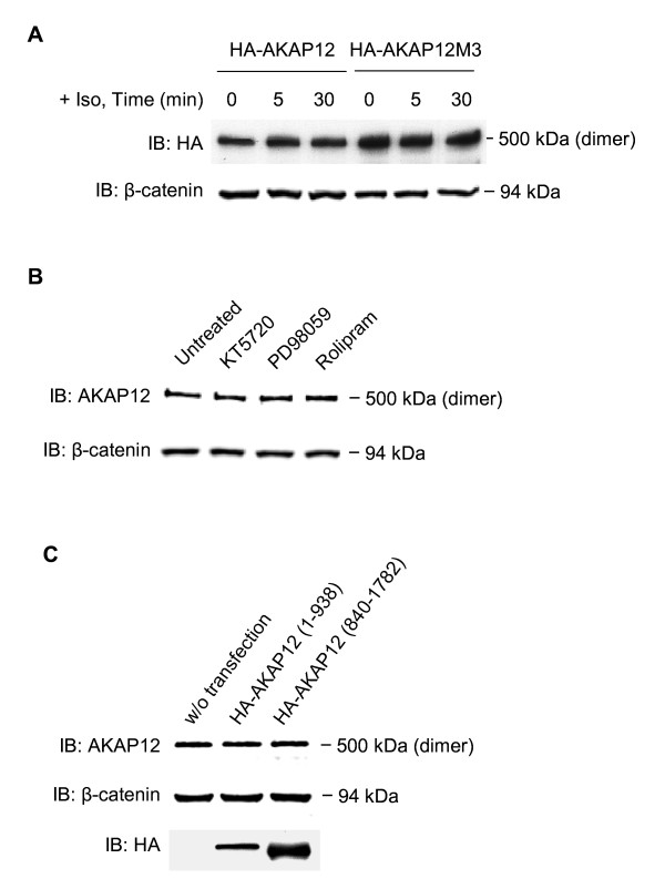
Figure 4
Oligomerization of AKAP12 in mammalian cells: elevation of intracellular cyclic AMP as well as inhibition of protein kinase A, MEK1/2, cyclic AMP phosphodiesterase 4, or expression of either N- or C-terminal fragments do not block AKAP oligomerization. (A) HEK293 cells were transfected with an expression vector harboring either HA-tagged full-length AKAP12 (HA-AKAP12) or an HA-tagged AKAP12 in which alanine substitution of protein kinase A phosphorylation sites (S627A, S696-698A, S772A) has been performed (AKAP12M3). Cells were treated with the beta-adrenergic agonist isoproterenol (10 μM) for 0, 5, or 30 min prior to harvesting the cells for lysis. Whole-cell lysates of transfected cells expressing either AKAP12 or AKAP12M3 were subjected to SDS-PAGE in the absence of 8 M urea. (B) HEK293 cells were either untreated or incubated with a chemical inhibitor for protein kinase A (KT5720, 1 μM), MEK1/2 (PD98059, 20 μM), or cyclic AMP phosphodiesterase 4 (Rolipram, 10 μM) for 45 min prior to cell lysis and subsequent analysis by SDS-PAGE. (C) HEK293 cells were either untransfected or transfected with an expression vector harboring the AKAP12 N-terminal (1-938) fragment or the C-terminal (840-1782) fragment. Whole-cell lysates of cells expressing either AKAP12 or one of the fragments subjected to SDS-PAGE in the absence of 8 M urea. The resolved proteins were transferred to PVDF membrane, subjected to immunoblotting, and made visible by staining with anti-HA or anti-AKAP12 antibodies. Beta-catenin was employed as a loading control for each lane, also identified by immunoblotting, stained with anti-beta-catenin antibodies. The results displayed are representative of at least three separate experiments performed on as many separate cell cultures.
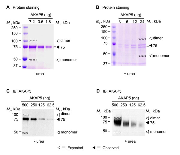
Figure 5
Oligomerization of AKAP5 expressed and purified from E. coli. His-tagged AKAP5 was expressed in E. coli. Affinity purified AKAP5 was resolved on either SDS-PAGE (A, C) or SDS-PAGE performed in the presence of 8 M urea (B, D). The gels were fixed and stained for protein with Coomassie Brilliant Blue (A, B) or the resolved proteins transferred to PVDF membrane and subjected to immunoblotting (C, D). Immunoblotting was performed using anti-AKAP5 antibody (C, D). The "◀" indicates the observed Mr on the gels and the "Δ" indicates the predicted Mr, calculated from the MW (47 kDa).
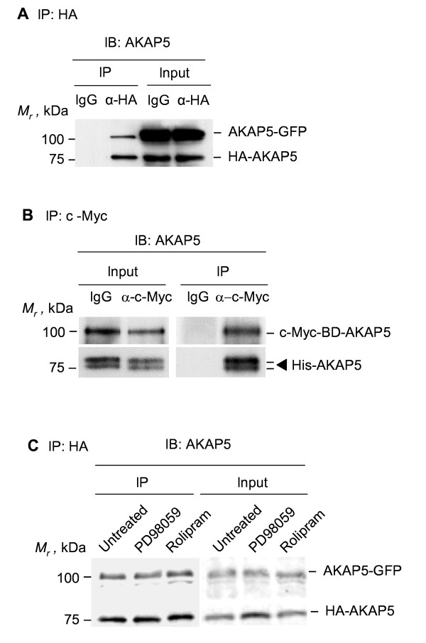
Figure 6
Oligomerization of AKAP5 in vivo and in vitro. (A) HEK293 cells stably expressing GFP-tagged AKAP5 were transiently transfected with HA-tagged AKAP5. Cell lysates were subjected to pull-downs mediated by IgG (control) or anti-HA antibodies (IP-HA) and the pulldowns subjected to SDS-PAGE, immunoblotting, and stained with anti-AKAP5 antibodies. Endogenous AKAP5 (expressed at <10% of that of exogenously expressed, tagged-AKAP) display a Mr identical with the HA-tagged version under these conditions. (B) c-Myc-tagged AKAP5 fused to amino acids 1-147 of the GAL4 DNA-binding domain (BD) was expressed in yeast. The c-Myc tagged AKAP5 was subjected to pull-down with anti-c-Myc antibody or control IgG. The immune complexes then were incubated with purified His-tagged AKAP5. The association of His-tagged AKAP5 with the c-Myc-tagged AKAP5 was detected by SDS-PAGE, immunoblotting, and staining of the blots with anti-AKAP5 antibodies. (C) HEK293 cells stably expressing GFP-tagged AKAP5 were transiently co-transfected with an expression vector harboring HA-tagged AKAP5. The cells were either untreated or incubated with a chemical inhibitor for either MEK1/2 (PD98059, 20 μM) or cyclic AMP phosphodiesterase 4 (Rolipram, 10 μM) for 45 min prior to cell lysis and analysis by SDS-PAGE. Pull-down of the HA-tagged AKAP5 was accomplished with anti-HA antibodies. The immune precipitates were subjected to SDS-PAGE and immunoblotting. The resolved, transferred protein blots were stained with anti-AKAP5 antibodies. The results displayed are representative of at least three separate experiments performed on as many separate cell cultures.
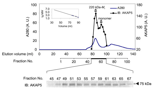
Figure 7
Oligomerization of AKAP5 expressed and purified from E. coil: analysis of higher-order assembly by steric-exclusion chromatography. His-tagged AKAP5 was expressed in E. coli. The AKAP5 was purified and then resolved by steric-exclusion chromatography performed on an AKTA FPLC fitted with HiPrep Sephacryl-S400 (16/60) column. The presence of supermolecular oligomers of AKAP5 was established by SDS-PAGE and immunoblotting of samples from the chromatography. The resolved, transferred proteins were stained with anti-AKAP5 antibodies. Marker protein mobilities were employed to establish the Mr of AKAP5 by elution position (inset). The results displayed are representative of two separate experiments performed on as many separate cell cultures.