
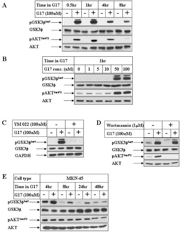
Figure 1
Effect of G17 on GSK3βSer9 phosphorylation in gastric cancer cells. (A) Confluent AGSE cells were treated in the absence (-) or presence (+) of 100 nM G17 in serum free media for the indicated periods of time. Equal amounts of total protein were fractionated by SDS-PAGE and subjected to Western Blot analysis utilizing antibodies against phospho-GSK3βSer9, total GSK3β, phospho-AKTSer473 and total AKT. (B) AGSE cells were treated with increasing concentrations of G17 for 1 hour followed by Western Blot analysis with the antibodies indicated. (C) & (D) Western Blot analysis of AGSE cells with the indicated antibodies, treated with 100 nM G17 for 1 hour following an overnight pretreatment with 100 nM YM 022 (C) or 1 μM Wortmannin (D). (E) MKN45 cells treated as in A were harvested at different time points following G17 treatment and analyzed by Western Blots utilizing the antibodies indicated.
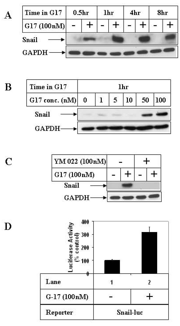
Figure 2
Effect of G17 on Snail expression in gastric cancer cells. (A) AGSE cells were treated as in 1A or (B) 1B above and subjected to Western Blot analysis utilizing antibodies against Snail and GAPDH (as control). (C) Western Blot analysis of cell extracts with the indicated antibodies, treated with 100 nM G17 for 1 hour, following an overnight pretreatment with 100 nM YM 022. (D) Subconfluent AGSE cells were transiently transfected with Snail-luciferase vector (Snail-luc) along with β-Gal vector (for normalization of transfection). Forty-eight hours after transfection, cells were treated overnight in the presence (+) or absence (-) of 100 nM G17, and luciferase and β-Gal assays were performed. The RLU/β-Gal values were represented as percent control, considering the untreated samples as 100%. Each transfection was performed in triplicate, and the data represent the mean ± SD of at least two independent experiments.
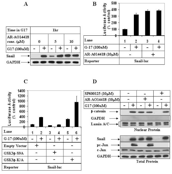
Figure 3
Effect of GSK3β inhibition on G17-induced Snail expression and β-catenin nuclear translocation. (A) AGSE cells were treated with (+) or without (-) 100 nM G17, following an overnight pretreatment with either none (lanes 1, 2), 5 μM (lanes 3, 4) or 10 μM (lanes 5, 6) AR-A014418. Western Blot analysis was then performed with the antibodies indicated. (B) Luciferase (with Snail-luc) and β-Gal assays were performed as in 2D following a 1 hour pretreatment with AR-A014418. (C) AGSE cells were co-transfected with Snail-luc and β-Gal vectors along with either Empty vector (lanes 1, 2), GSK3β-S9A mutant vector (lanes 3, 4) or GSK3β-K/A mutant vector (lanes 5, 6). Luciferase and β-Gal assays were performed after G17 treatment as in 2D. Each transfection (3B, 3C) was performed in triplicate, and the data represent the mean ± SD of at least two independent experiments. (D) Upper Panel: Confluent AGSE cells were treated with G17 for 8 hours after an overnight pretreatment with none (lanes 1, 2), or AR-A014418 (lanes 3, 4) or SP600125 (lanes 5, 6). At the end of treatment, nuclear protein was isolated and subjected to Western Blot analysis with antibodies against β-catenin, GAPDH (cytoplasmic protein) or Lamin A/C (nuclear protein). Lower Panel: Cells were pretreated as in the upper panel, followed by 1 hour G17 treatment and Western Blot analysis.
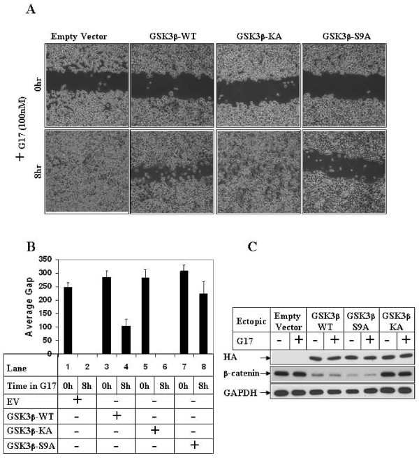
Figure 4
Effect of overexpression of GSK3β on G17-induced migration. (A). Subconfluent AGSE cells were transiently transfected with Empty Vector, GSK3β-WT, GSK3β-KA mutant or GSK3β-S9A mutant vectors. The cells were wounded linearly 48 hours post-transfection and, after an overnight recovery following wounding, they were treated with G17 and pictures obtained at the indicated times. (B) AGSE cells were transfected as in 4A followed by G17 treatment and wound healing assay. The distance of migration of the wounded edges for each time point were measured at several places and the average distance was represented by bar diagrams as "Average Gap". (C) AGSE cells transfected in A and treated with G17 were analyzed for protein expression. Western Blot analysis was performed with an HA.11 antibody to detect ectopic HA-tagged GSK3β proteins and with β-catenin and GAPDH antibodies to detect the corresponding endogenous proteins.
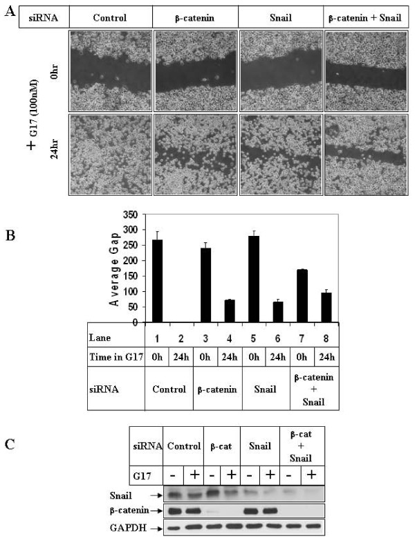
Figure 5
Effect of knockdown of β-catenin and Snail expression on G17-induced migration. (A). Subconfluent AGSE cells were transiently transfected with 100 nM each of either control-siRNA, or β-catenin-siRNA, or Snail-siRNA or a combination of β-catenin and Snail siRNA. They were wounded 48 hours post-transfection and treated with G17 and pictures were obtained as described under 4A. (B) The distance of migration of the wounded edges were measured as in 4B and represented as "Average Gap". (C) AGSE cells transfected in A and treated with G17 were analyzed by Western Blot analysis utilizing the indicated antibodies.
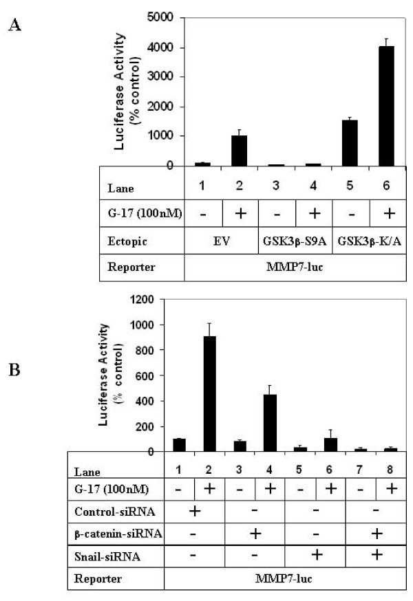
Figure 6
Effect of modulation of GSK3β, β-catenin and Snail pathways on G17-induced MMP7 transcription. (A). Subconfluent AGSE cells were co-transfected with MMP7-luciferase and β-Gal vectors along with either EV (lanes 1, 2), GSK3β-S9A (lanes 3, 4) or GSK3β-K/A (lanes 5, 6) mutants. Luciferase and β-Gal assays were performed after G17 treatment as described under 2D. (B) AGSE cells were transfected as in A along with either control-siRNA (lanes 1, 2), β-catenin-siRNA (lanes 3, 4), Snail-siRNA (lanes 5, 6) or a combination of β-catenin and Snail-siRNA (lanes 7, 8). G17 treatment was performed as described under 2D, followed by luciferase and β-Gal assays. Each transfection (A and B) was performed in triplicate, and the data represent the mean ± SD of at least two independent experiments.
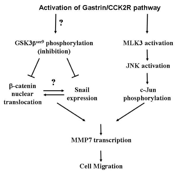
Figure 7
Model representing the signaling pathway of G17-induced migration. Stimulation of G17/CCK2R pathway leads to activation of two separate signaling axes: (i) an activation of MLK3/JNK1 axis, which via activation of its downstream transcription factor c-Jun induces MMP7 transcription leading to increased migration; (ii) an induction of GSK3βSer9 phosphorylation (inhibition of axis) via a PI3Kinase-independent (and yet unknown) mechanism. This leads to an increase in Snail protein expression and β-catenin nuclear translocation, combination of which lead to increased MMP7 transcription and cell migration.