
Figure 1
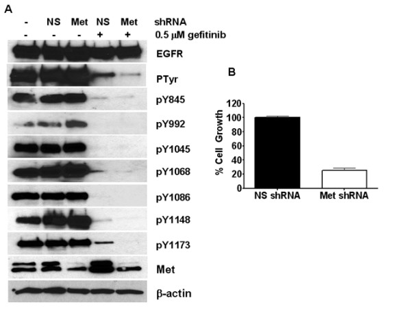
Met expression mediates EGFR tyrosine phosphorylation and proliferation in the absence of EGFR kinase activity. (A) SUM229 cells were infected with lentiviral particles containing Met shRNA or a non-silencing shRNA for 72 hrs. Cells were treated with gefitinib for the last hour of infection at 0.5 μM, cells were lysed, and lysates were immunoprecipitated with anti-EGFR. Immunoprecipitates were immunoblotted with anti-EGFR, anti-Ptyr, and the indicated phospho-specific sites on the EGFR. Corresponding whole cell lysates were immunoblotted with anti-Met and β-actin as a loaded control. Through quantification, EGFR phosphorylation was decreased by 53% in the gefitinib treated cells and 88% in the gefitinib with Met knocked down cells. The two bands in the Met immunoblot represent processed and pro-forms of Met. (B) SUM229 cells were infected with lentiviral particles containing Met shRNA in the presence of gefitinib or a non-silencing control for seven days in the presence of puromycin to select for infected cells. Cells were counted using a Coulter Counter and the day 8 values were graphed with the error bars representing SEM.
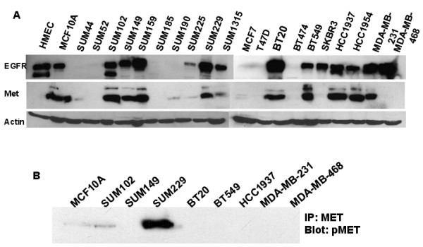
Figure 2
Met is not amplified, but is overexpressed at the protein level. (A) Lysates from the indicated breast cancer cell lines was prepared and 100 ug of protein was separated by SDS-PAGE, transferred, and immunoblotted with anti-EGFR, anti-Met, and anti-β-actin. (B) Lysates from the Met expressing breast cancer cell lines were immunoprecipitated with anti-Met antibodies. Immunoprecipitates were separated by SDS-PAGE, transferred, and immunoblotted with anti-pMet antibodies.
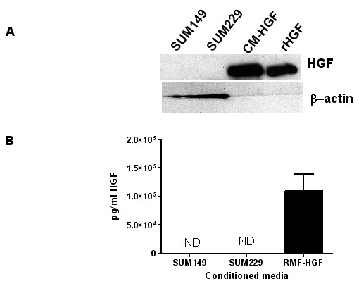
Figure 3
HGF is not expressed in SUM229 breast cancer cells. (A) Lysates from SUM149 and SUM229 and conditioned media from RMF-HGF cells were immunoblotted with anti-HGFα. Recombinant HGF was used as a positive control. (B) Conditioned media was collected from confluent SUM149, SUM229, and RMF-HGF cell cultures. Diluted conditioned media was analyzed for HGF expression using ELISA with RMF-HGF as a positive control. The amount of HGF produced from the SUM149 and SUM229 lysates was undetectable.
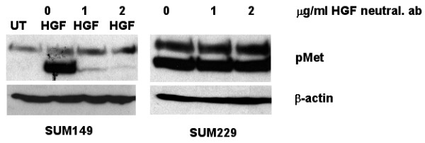
Figure 4
Met phosphorylation occurs independent of ligand. SUM149 and SUM229 cells were treated with increasing concentrations of anti-HGF neutralizing antibody. The SUM149 cells were stimulated with 50 ng/ml HGF to induce Met phosphorylation. Whole cell lysates were immunoblotted with anti-phospho-Met.
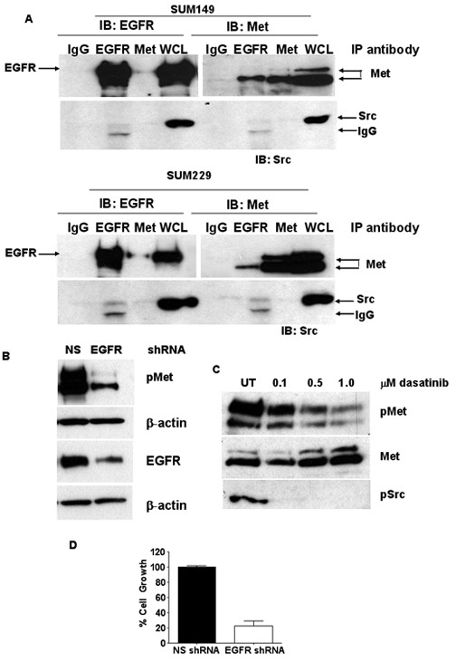
Figure 5
EGFR and Met association independent of EGFR kinase activity. (A) SUM149 or SUM229 cells were treated with 0.5 μM gefitinib or DMSO control for 30 min. Cells were lysed and immunoprecipitated with anti-EGFR, anti-Met, and anti-IgG. Whole cell lysates were used as a control. Immunoprecipitates were separated by SDS-PAGE, transferred, and immunoblotted using anti-EGFR, anti-Met, or anti-c-Src antibodies. (B) SUM229 cells were incubated with EGFR shRNA lentiviral particles for 72 hrs. Lysates were prepared and separated by SDS-PAGE. Membranes were immunoblotted with anti-pMet, anti-Met, anti-EGFR, and β-actin. (C) SUM229 cells were treated with increasing concentrations of dasatinib for 2 hrs. Lysates were prepared and immunoblotted with anti-pMet, anti-Met, and anti-pSrc antibodies. (D) SUM229 cells were incubated with EGFR shRNA lentiviral particles for 24 hrs and then treated with puromycin to select for virus expressing cells. After the puromycin was added for 24 hrs, the cells were grown for 7 days at which point cell numbers were determined via Coulter counting.
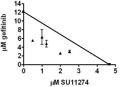
Figure 6
EGFR and Met inhibitor synergy in SUM229 cells. SUM229 cells were treated with various concentrations of gefitinib and SU11274 and the IC50 of gefitinib was calculated for each concentration of SU11274. A line was drawn between the IC50 for gefitinib (y-axis; 12.2 μM) and SU11274 (x-axis; 4.8 μM). The calculated IC50 values for gefitinib with SU11274 combination treatment were plotted. Points falling below the line represent synergistic drug interactions.