
Figure 1
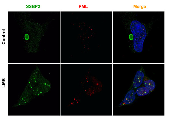
SSBP2 is localized in the nucleus of HEK293 cells in response to CRM1-export pathway inhibition. Exponentially growing HEK293 cells were treated with vehicle (upper panel) or 10nM LMB (lower panel) for 3 hrs. prior to fixation and staining with antibodies to SSBP2 [7] or PML (red). Cells were co-stained with DAPI and the images obtained by deconvolution algorithms are shown. In control cells SSBP2 localizes to the large aggresome, while PML is in distinct nuclear bodies (upper panel). When nuclear export is blocked by LMB treatment, SSBP2 and PML localize to nuclear structures (lower panel).
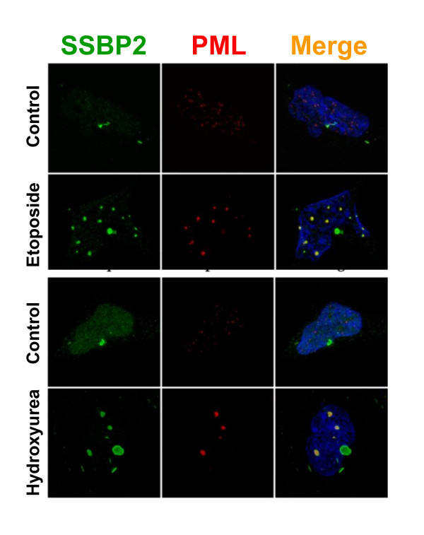
Figure 2
SSBP2 and PML colocalize in PML-NBs in response to DNA damage in HEK293 cells. Cells were treated with 1uM etoposide (A) or 0.2 uM hydroxy urea (B) for 24 hours and stained with antibodies to SSBP2 [7] and PML (red). Merged images are shown on the far right. Note the colocalization of SSBP2 and PML only in the PML-NBs in response to DNA damage.
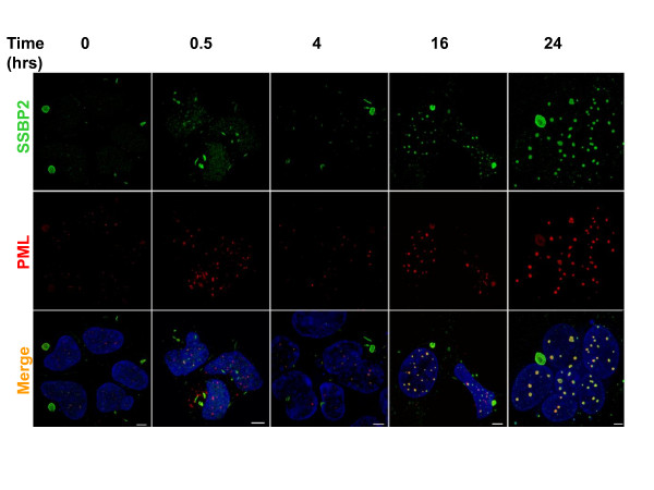
Figure 3
Kinetics of PML-NB formation in HEK293 cells in response to radiation. Cultures were exposed to 10gy irradiation and allowed to recover in media at 37°C for 0.5-24 hrs as indicated. Images from SSBP2[7], PML(red) stains and overlay of both (yellow) are depicted. Bars denote 10uM.
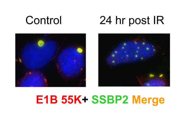
Figure 4
E1B 55K and SSBP2 are redirected to nuclear bodies after irradiation. Merged image of E1B55K (red) and SSBP2 [7] shows localization to aggresome in the control HEK293 in contrast to the nuclear bodies 24 hrs after irradiation.
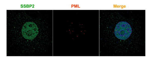
Figure 5
SSBP2 does not localize to PML-NBs in response to radiation in MCF7 cells. Cells were exposed to 10gy ionizing radiation and allowed to recover for 7 h. SSBP2 localizes to distinct nuclear structures and cytoplasmic structures. No colocalization with PML is detected.