
Figure 1
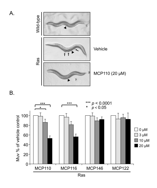
MCP110 and MCP116 but not MCP146 inhibit the Ras/LET-60-induced Muv phenotype. A. Representative images of untreated wild-type animals (WT phenotype with normal vulva (arrowhead) but no ventral protrusions); animals harboring constitutively activated Ras treated with vehicle only (Muv phenotype; arrows mark ventral protrusions formed by pseudovulvae); the same strain following treatment with MCP110 (20 μM) (WT phenotype, as indicated by lack of ventral protrusions). B. Animals harboring constitutively activated Ras as in Panel A were treated with either vehicle (DMSO) or MCP110, MCP116, MCP146 or negative control MCP122 (3, 10 or 20 μM; higher concentrations precipitated out of solution). The Y-axis indicates the percentage of MCP-treated animals with a Muv phenotype, normalized to vehicle-treated animals. Data were analyzed by three-way ANOVA. (***) and (*) indicate p values of < 0.0001 and < 0.05, respectively.
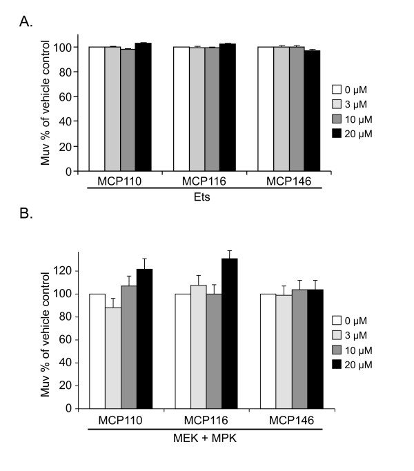
Figure 2
MCP110 and derivatives do not inhibit the Muv phenotype induced by activation of the Ras-Raf-MAPK pathway downstream of Raf. Animals were treated, data were analyzed and results are presented as described in the legend to Figure 1B. A. Ets and B. activated MEK/ERK.
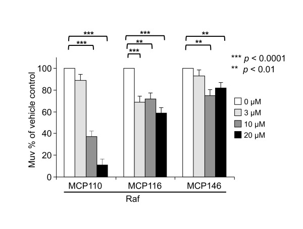
Figure 3
MCP110 inhibits the Raf-AA Muv phenotype. Animals expressing a constitutively activated Raf/LIN-45 protein were treated with vehicle (DMSO) or MCP110 as in Figure 1B. Bar graphs show the percentage of MCP-treated animals with a Muv phenotype, normalized to vehicle-treated animals. (***) and (**) indicate p-values of < 0.0001 and < 0.01, respectively.
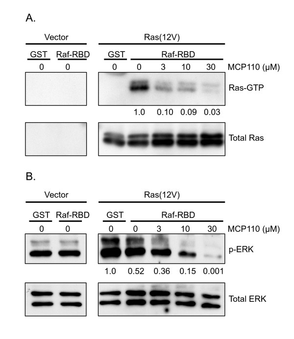
Figure 4
MCP110 inhibits Ras/Raf interaction and signaling to ERK in mammalian cells. A. Pulldown assay for Ras-Raf interaction. Pulldowns of active Ras bound to Raf-RBD were done using GST-Raf-RBD as described in Methods. NIH 3T3 cells were treated with vehicle or MCP110 (3, 10 and 30 μM) before and after lysis. Ras was detected by immunoblotting with anti-Ras antibody. Both Raf-RBD-bound Ras (Ras-GTP, upper panel) and total Ras in the lysates (lower panel) are shown. MCP110 disrupted the Ras/Raf interaction in a dose-dependent manner; numbers shown below panels indicate quantitation of Ras pulldown by densitometry, normalized to vehicle control. B. Western blot analysis for active, phospho-ERK1/2. The same lysates from cells depicted in panel A above were immunoblotted for phospho-ERK1/2 (P-ERK, upper panel) and for total ERK1/2 (total ERK, lower panel). Numbers shown below PERK panel indicate quantitation by densitometry, normalized to vehicle control. MCP110 decreased phospho-ERK1/2 in a dose-dependent manner.
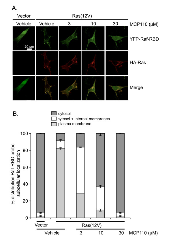
Figure 5
MCP110 impairs recruitment of Raf-RBD to subcellular locations of active Ras. A. Recruitment of YFP-Raf-RBD to sites where Ras is localized, as detected by fluorescence microscopy. NIH3T3 cells transiently expressing both HA-tagged active Ras and YFP-Raf-RBD were treated with vehicle or MCP110. Shown are representative images of cells quantitated in Panel B below. In the absence of active Ras (v.o.), YFP-Raf-RBD (green) was localized diffusely throughout the cytoplasm and nucleus, whereas in the presence of active Ras, the Raf-RBD probe was recruited to the plasma membrane and, like Ras (red), was nuclear-excluded ("vehicle" panels). Increasing concentrations of MCP110 increasingly shifted the YFP-Raf-RBD probe from the plasma membrane to the cytosol and to internal membranes and finally to both cytosol and nucleus, whereas Ras remained membrane-associated and nuclear-excluded. These results indicate dose-dependent disruption of the Ras/Raf-RBD interaction by MCP110. B. Quantification of the distribution of YFP-Raf-RBD subcellular localization. Cells treated and analyzed as described in Methods and depicted qualitatively in Panel C were binned according to whether the YFP-Raf-RBD probe accumulated primarily in the cytosol, or cytosol + internal membranes, or at the plasma membrane and was nuclear-excluded. MCP110 dose-dependently disrupts the ability of Ras to recruit Raf-RBD.