
Figure 1
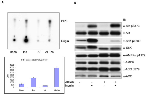
Effect of AICAR on insulin-stimulated phosphorylation of the PI3K pathway. 3T3-F442a preadipocytes were differentiated into adipocytes. A. The cells were treated with or without AICAR (AI) (1 mM, 2 h), followed by insulin (Ins) (100 nM, 15 min) and equal amounts of cell lysates were immunoprecipitated with antibodies against IRS1 and assayed on the associated PI3K activity. The radiolabeled PIP3 lipids were resolved on thin layer chromatography and exposed to X-ray film. The autoradiogram represents one of a triplicate experiment. The PIP3 spots were excised and counted in liquid scintillation. The graph denotes the average CPM of labeled PIP3 (n = 3, mean ± SD). B. In parallel to A, protein samples were run onto SDS-PAGE and blotted with antibodies, as indicated.
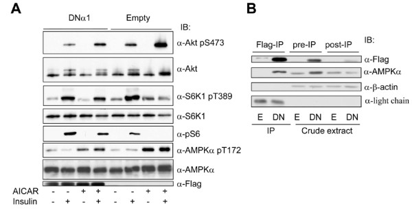
Figure 2
Dominant negative mutant of AMPK α1 subunit suppresses the effect of AICAR. 3T3-F44a preadipocytes were stably infected with lentivirus expressing a dominant negative mutant of AMPK α1 catalytic subunit (DNα1) tagged with Flag epitope or a control virus (Empty). A. The cells were treated with AICAR and insulin, as described for Figure 1, and assayed for immunoblot with antibodies, as indicated. B. Expression of DNα1 diminishes the endogenous counterpart. The cell extracts containing empty vector (E) or the AMPK mutant (DN) were immunoprecipitated (IP) with anti-flag antibody and equal amounts of pre-IP and post-IP extracts were blotted with anti-flag and anti-AMPK α1 antibodies, respectively. β-actin blot was used as an internal control.
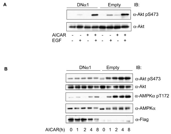
Figure 3
AMPK promotes Akt phosphorylation independent of IRS1. A. 3T3-F44a preadipocytes were treated with AICAR (1 mM, 2 h), followed by EGF (10 ng/ml, 15 min) and the cell extracts blotted with antibodies, as indicated. B. 3T3-F44a preadipocytes containing DNα1 or the empty viral vector were incubated with AICAR (1 mM) for different time, and the cell extracts blotted with antibodies, as indicated.
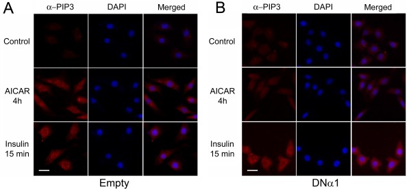
Figure 4
AMPK activation stimulates production of PIP3. 3T3-F44a preadipocytes bearing empty lentiviral vector (A) or the dominant negative mutant of AMPKα1 subunit (B) were treated with AICAR (1 mM, 4 h) or insulin (100 nM, 15 min). Cells were immunostained with anti-PIP3 monoclonal IgM antibody and counter-stained with DAPI for visualization of cell nuclei. Cells with empty viral vector accumulated PIP3 when treated with AICAR, as well as insulin (A). The AICAR induced PIP3 accumulation was not seen in the cells containing the AMPK mutant, whereas the effect of insulin was not affected (B). Scale bar: 63.4 μm.
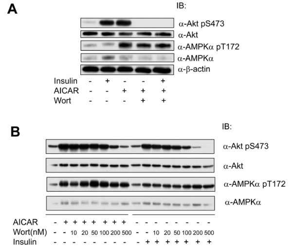
Figure 5
Akt Activation by AICAR is suppressed by Wortmannin. A. 3T3-F44a preadipocytes were incubated with Wortminnin (Wort, 0.5 μM, 30 min), followed by AICAR (1 mM, 4 h) or insulin (100 nM, 15 min). The cell extracts were blotted with antibodies, as indicated. B. 3T3-F44a preadipocytes were treated with Wortmannin at different doses, as indicated, for 30 min and followed by treatment with AICAR or insulin, as described for A. Cell extracts were blotted with antibodies, as indicated.
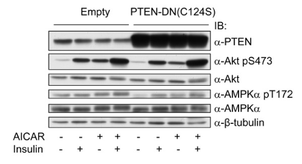
Figure 6
Inhibition of PTEN does not affect AMPK-induced activation of Akt. 3T3-F44a preadipocytes stably infected with the dominant negative PTEN (C124S) were treated with AICAR, insulin or together, as described for Figure 1, and the cell extracts were blotted with antibodies, as indicated.