
Figure 1
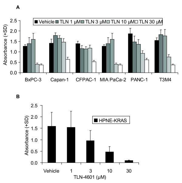
TLN-4601 reduces contact-dependent proliferation in PDAC (A) and Ras-transformed immortalized duct derived cells (B). Cells were treated for 72 h with vehicle or various concentrations of TLN-4601 prior to the addition of the MTT reagent. Data represent the average of quadruplicate wells + SD of three independent experiments.
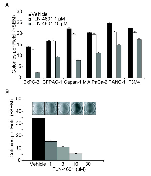
Figure 2
TLN-4601 reduces contact-independent proliferation in PDAC (A) and Ras-transformed immortalized duct derived cells (B). Cells were seeded into soft agar containing vehicle or various concentrations of TLN-4601. Bars represent the mean colonies per field, averaged from five fields per well, triplicate wells + SEM, n = 3 independent experiments. Insets are representative wells.
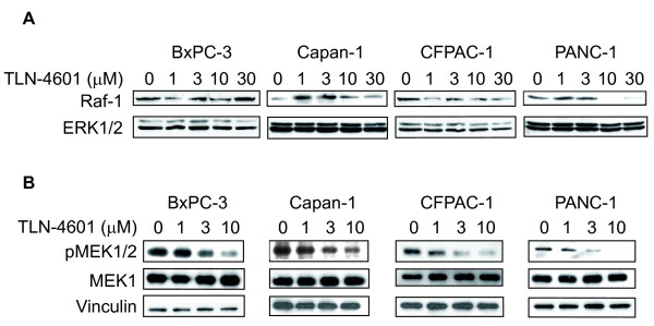
Figure 3
TLN-4601 inhibits the K-Ras downstream signaling cascade in PDAC cells. Exponentially growing cells were treated with increasing concentrations of TLN-4601. After overnight exposure, cells were lysed and cellular extracts (30 μg protein) were separated on a 10% SDS-PAGE gel and transferred to nitrocellulose membranes. (A) Membranes were sequentially probed with Raf-1 and ERK1/2 (p44/42 MAP kinase) or (B) phospho-MEK1/2 (Ser217/221), MEK1 and vinculin (loading control). Blots are representatives of three independent experiments.
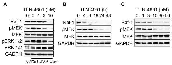
Figure 4
TLN-4601 reduces K-Ras related signaling in EGF- (A) and serum-stimulated MIA PaCa-2 cells (B,C). Cells, treated with either escalating doses (A, C) or increasing time (B) of TLN-4601, were harvested and lysed for western blot analysis. GAPDH was used as a loading control.
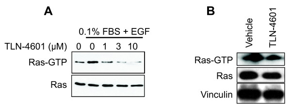
Figure 5
Dose-dependent reduction of K-Ras GTP in MIA PaCa-2 (A) and immortalized K-Ras12D expressing cells (B). Exponentially growing MIA PaCa-2 cells serum-starved starved for 9 h in DMEM with 0.1% FBS and then treated for 18 h with 0 (DMSO control), 1, 3 or 10 μM of TLN-4601. At the end of the treatment, cells were stimulated with EGF (100 mg/ml) for 5 min. HPNE-KRAS cells were grown in normal serum condition and treated for 18 h with 10 μM of TLN-4601. Pull-down analyses and western blot analyses with K-Ras-specific antibody were done to determine the level of activated and total K-Ras protein expression. Data are representative of one to three independent experiments. Vinculin was used as a loading control.
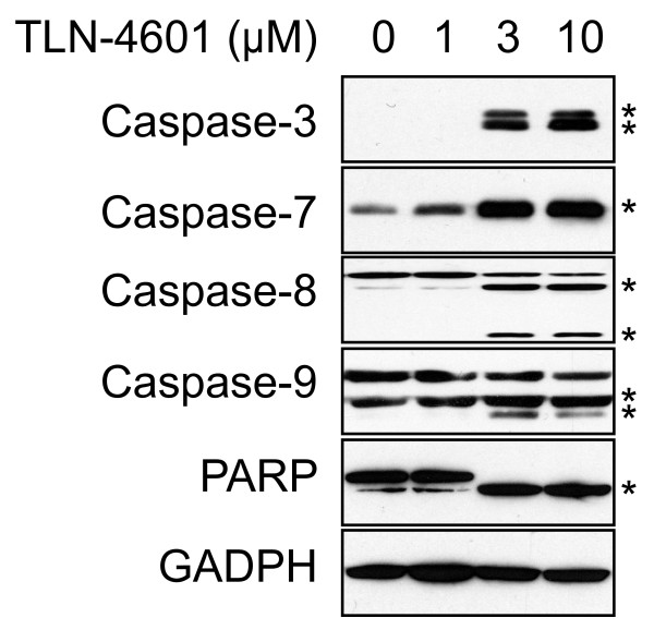
Figure 6
TLN-4601 activates the apoptosis cascade in MIA PaCa-2 cells. Lysate from TLN-4601-treated cells were separated by SDS-PAGE and probed for caspases and PARP activation and cleavage. GAPDH was used as a loading control. * Cleaved forms of different caspases and PARP
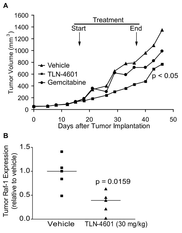
Figure 7
TLN-4601 inhibits tumor growth and K-Ras signaling in vivo. MIA PaCa-2 xenograft-bearing nude mice were treated with TLN-4601 (30 mg/kg s.c., once a day Monday through Friday for three consecutive weeks) or gemcitabine (60 mg/kg i.p., twice a week for 4 weeks). Control group received 5 ml/kg of drug-free vehicle (15% PS80/5% PEG 400/5% EtOH/80% D5W) s.c., once a day Monday through Friday for three consecutive weeks. (A). Tumors were excised, lysed, and probed for Raf-1 protein expression (B); horizontal lines mark the mean of n = 5 mice.