
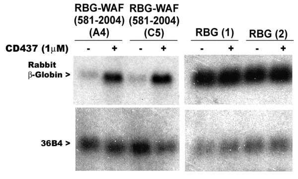
Figure 1
CD437 regulation of chimeric rabbit β-globin-p21WAF1/CIP1 mRNA. Two independent sublines derived from transfection of RGB-WAF (581-2004) plasmid [31.4(A4) and 31.4(C5)] or RBG plasmid [29.6(1) and 29.6(2)] were grown either in the absence (-) or presence (+) of 1 μM CD437 for 48 hours. Total RNAs were prepared and expression of RBG transcripts analyzed by northern blot hybridization as described [12]. Levels of RNA loading in each lane were assessed by signals from hybridization with ribosomal phosphoprotein 36B4 cDNA [18]
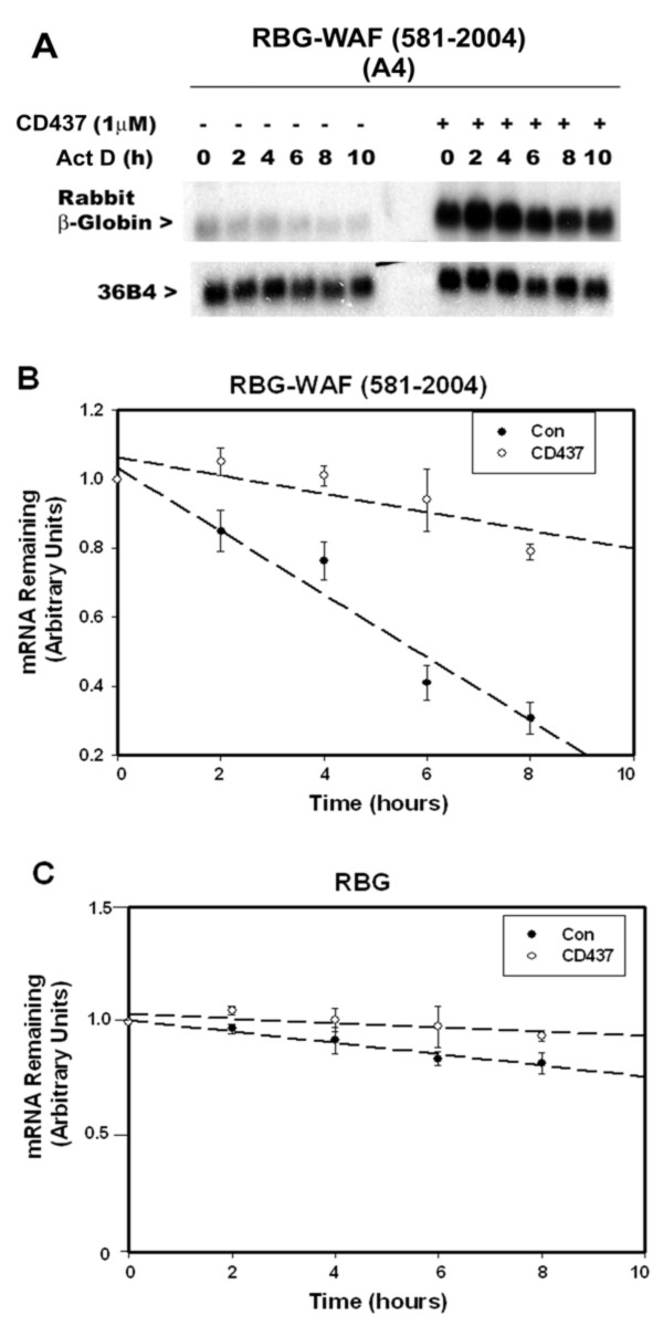
Figure 2
Presence of p21WAF1/CIP1 3'-UTR causes destabilization of RBG mRNA, while CD437 treatments enhance stability of chimeric RBG-WAF transcripts. MDA-MB-468 sublines derived from stable transfection of RBG-WAF (581-2004) plasmid (Panel A) or RBG plasmid (not shown) were cultured in the absence (-) or presence (+) of CD437 for 48 hours and exposed to transcriptional inhibitor ACT D for noted times. The preparation of RNA and northern blot hybridization were as described in the legend to figure 1. Panels B and C show plots of the decay of RBG transcripts in either the presence or absence of CD437. The RBG mRNA levels at time 0 in either the absence or presence of CD437 were arbitrarily defined as 1. Data in panels B and C represent mean of four and two independent sublines, respectively, for each time point. Error bars represent the standard error of the mean.
Table 1
CD437-responsive sequences of p21WAF1/CIP1 3'-UTR
| Construct ID | Plasmid Name | Positions of p21WAF1/CIP1 3'-UTR insert [4] | CD437 Responsiveness |
|---|---|---|---|
| 29.6 | RBG | ------- | ------- |
| 31.4 | RBG-WAF (581-2004) | +581 to +2004 | Yes |
| 30.2 | RBG-WAF (1006-2004) | +1006 to +2004 | Yes |
| 32.8 | RBG-WAF (1498-2004) | +1498 to +2004 | Yes |
| 33.7 | RBG-WAF (1645-2004) | +1645 to +2004 | Yes |
| 36.4 | RBG-WAF (1795-2004) | +1795 to +2004 | No |
| 37.4 | RBG-WAF (1900-2004) | +1900 to +2004 | No |
| 38.1 | RBG-WAF (1498-1645) | +1498 to +1645 | Yes |
| 49.1 | RBG-WAF (1540-1700) | +1540 to +1700 | Yes |
| 50.1 | WAF (1540-1700)-RBG | +1540 to +1700 | Yes |
| 60.3 | RBG-WAF (581-1498) | +581 to +1498 | Yes |
| 61.2 | RBG-WAF (581-1195) | +581 to +1195 | No |
| 62.3 | RBG-WAF (581-1011) | +581 to +1011 | No |
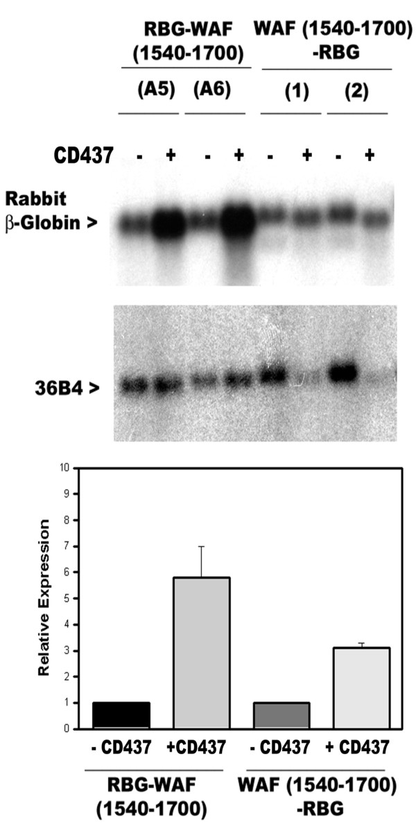
Figure 3
CD437 targets 3'-UTR sequences of p21WAF1/CIP1 mRNA independent of their location in the chimeric RBG transcripts. (A) Two independent sublines derived from stable transfection of each of the RBG-WAF (1540-1700) plasmid (labelled as A5 and A6) and WAF (1540-1700)-RBG plasmid (labeled as 1, 2) were grown either in the absence (-) or presence (+) of 1 μM CD437 for 48 hours. The cellular RNAs were prepared and northern blot hybridizations carried out as described in the legend to figure 1. (B) Histogram showing relative expression of chimeric RBG-WAF transcripts in control vs CD437-treated sublines in panel A. Northern blots of panel A were quantitated by soft laser densitometry. Columns represent mRNA levels relative to the levels of mRNAs present in the untreated control lanes, which were arbitrarily defined as 1. The error bars represent the standard error of the mean.
Table 2
Plasmid constructs for in vitro transcription
| Construct I.D. | Plasmid Name | Positions of the UTR inserts | Gene [ref] |
|---|---|---|---|
| WAF 14.2 | pBKS-WAF (518-2110) | 3'-UTR +581 to +2110 | P21WAF1/CIP1 [1] |
| WAF 52.1 | pBKS-WAF (1540-1710) | 3'-UTR +1540 to +1700 | P21WAF1/CIP1 [1] |
| WAF 55.1 | pBKS-WAF (1540-1585) | 3'-UTR +1540 to +1585 | P21WAF1/CIP1 [1] |
| WAF 56.5 | pBKS-WAF (1575-1620) | 3'-UTR +1575 to +1620 | P21WAF1/CIP1 [1] |
| WAF 57.8 | pBKS-WAF (1610-1655) | 3'-UTR +1610 to +1655 | P21WAF1/CIP1 [1] |
| WAF 58.3 | pBKS-WAF (1645-1690) | 3'-UTR +1645 to +1690 | P21WAF1/CIP1 [1] |
| GADD 12.4 | pBKS-GADD (10-55) | 5'-UTR +10 to +55 | GADD45 [13] |
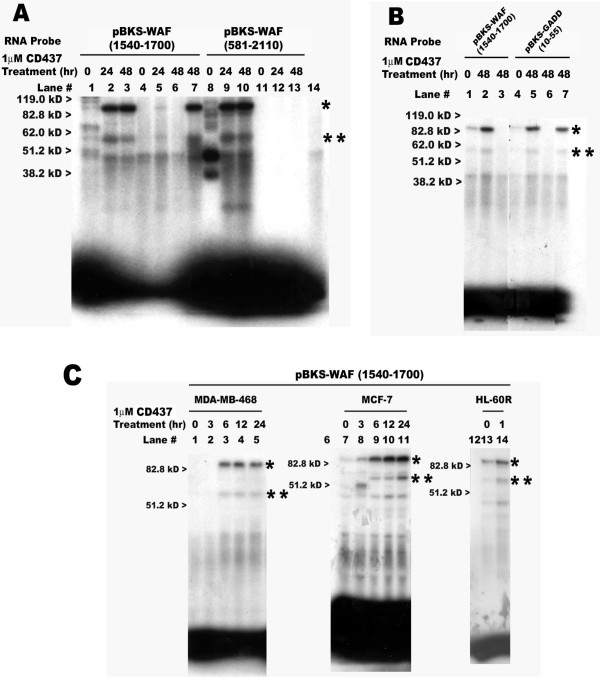
Figure 4
Multiple cell cytoplasmic proteins bind to radiolabelled p21WAF1/CIP1 3'-UTR and GADD45 5'-UTR sense strand riboprobes. Transcription and labeling of the indicated probes as well as the in vitro binding reactions were as described in the Methods. The probes utilized and treatment times with CD437 (hr) are indicated above each lane in the respective panels. The cytoplasmic protein extracts were prepared from untreated (lanes marked 0 hours) or CD437-treated MDA-MB-468 (panels A-C), MCF-7 (panel C) and HL-60R (panel C) cells. Panel A, the protein extracts in lanes 4-6 and11-13 were pre-incubated with 200-fold excess of the respective unlabeled probe RNAs, while the protein extracts in lane 7 were pre-incubated with 200-fold excess of the unlabeled 3'-UTR of c-myc RNA followed by incubation with the indicated labeled probe RNAs. Probe 14.2 RNA is loaded in lane 14. Panel B, the protein extracts in lanes 3 and 6 were pre-incubated with 200-fold excess of the respective unlabeled probe RNAs, while the protein extracts in lane 7 were pre-incubated with 200-fold excess of the unlabeled 3'-UTR of c-myc RNA followed by incubation with the indicated labeled probe RNAs. Panel C, lanes 6, 12 represent the labeled probe only. The approximate migration of molecular weight standards is marked on the left side of respective panel, while the * and ** on the right side of each panel denote locations of the putative 85 kD and 55 kD complexes, respectively.
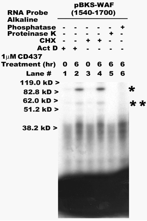
Figure 5
CD437-dependent RNA-protein interactions are independent of nascent protein synthesis or transcription but are phosphorylation dependent. The preparation of cytoplasmic protein extracts from untreated (lanes 0 hour) or CD437-treated (lanes 2, and 4-6, 6 hours) MDA-MB-468 HBC cells, and transcription and labeling of the indicated probe as well as in vitro binding reactions were as described in the methods. The cytoplasmic extracts were prepared from HBC cells pre-treated with protein synthesis inhibitor CHX (lanes 1, 2) or transcriptional inhibitor ActD (lanes 3, 4) followed by treatments with CD437 (time and dose indicated). In addition, cytoplasmic protein extracts derived from CD437-treated HBC cells were pretreated with alkaline phosphatase (lane 6) or proteinase K (lane 5) prior to binding with the indicated probe. The approximate migration of molecular weight standards is marked on the left side, whereas the * and ** on the right side of the panel denote locations of the putative 85 kD and 55 kD complexes, respectively.
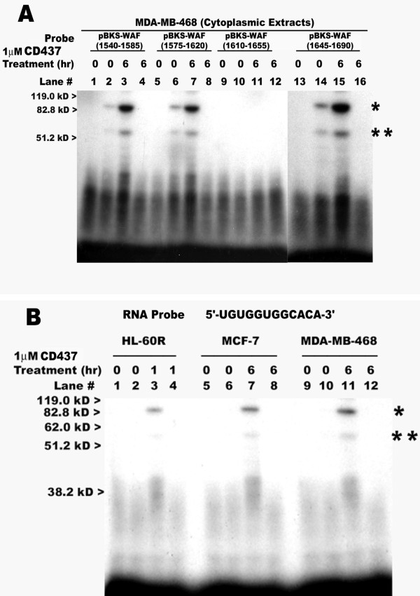
Figure 6
Multiple CD437-responsive sequence elements exist within the 3'-UTR of p21WAF1/CIP1. Transcription and labeling of the indicated probes and binding reactions were as described in the Methods. The probes utilized and treatment times with CD437 (hours, hr) are indicated in the respective panels. In panel B, the probe sequence of the 12 nt RNA oligonucleotide is shown. The cytoplasmic protein extracts were prepared from untreated (lanes marked 0) or CD437-treated MDA-MB-468 (panels A, B), MCF-7 (panel B) and HL-60R (panel B) cells. The respective probes only were loaded in lanes 1, 5, 9, and 13. The protein extracts in lanes 4, 8, 12, and 16 were pre-incubated with 200-fold excess of the unlabeled RNA derived from pBKS-WAF (1540-1700) plasmid (table 2), followed by incubation with the indicated labeled probe. The approximate migration of molecular weight standards is marked on the left side of respective panel, while the * and ** on the right side of each panel denote locations of the putative 85 kD and 55 kD complexes, respectively.