
Figure 1
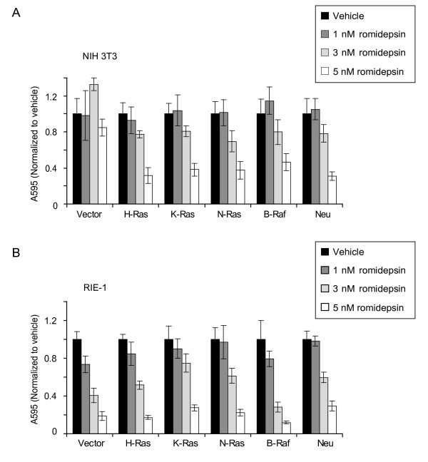
Romidepsin inhibits the anchorage-dependent growth of Ras-transformed NIH 3T3 and RIE-1 cells. NIH 3T3 cells (A) and RIE-1 cells (B) stably-infected with the empty pBabe-puro vector or encoding constitutively active human H-Ras(G12V), K-Ras(G12V), N-Ras(G12D), or B-Raf(V600E), or rat ErbB2/Neu(V664E) were seeded on day 0. On day 1, cells were treated with complete growth medium supplemented with DMSO (Vehicle), or 1, 3, or 5 nM romidepsin. After 72 h of romidepin treatment (day 3), cell viability was measured using the MTT assay. Data shown are the average ± SD of eight replicate wells and are representative of three independent experiments.
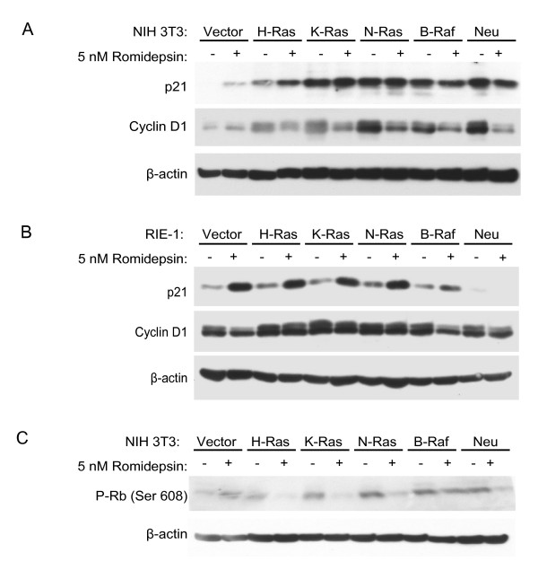
Figure 2
Romidepsin affects expression of cell cycle regulators. NIH 3T3 (A, C) or RIE-1 (B, D) cells stably expressing the indicated proteins were treated with DMSO (Vehicle) or the indicated concentration of romidepsin for 24 h. Cell lysates were analyzed by western blotting with antibodies to cyclin D1, p21, phospho-S608-Rb, and β-actin (loading control). Data shown are representative of at least two independent experiments.
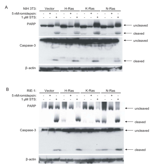
Figure 3
Romidepsin does not induce cleavage of caspase-3 or PARP. NIH 3T3 cells (A) and RIE-1 cells (B) stably infected with pBabe.puro vector or encoding H-Ras(G12V), K-Ras(G12V), or N-Ras(G12D) were treated with 5 nM romidepsin for 72 h or 1 μM staurosporine (STS) for 24 h. Cell lysates were analyzed by western blotting with antibodies to PARP, caspase-3, and β-actin (loading control). Data shown are representative of at least three independent experiments.
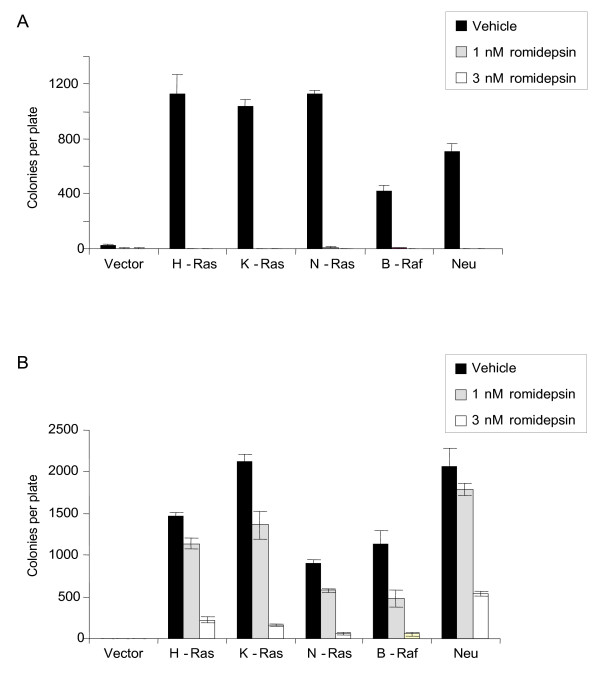
Figure 4
Romidepsin inhibits anchorage-independent growth of transformed NIH 3T3 and RIE-1 cells. NIH 3T3 cells (A) and RIE-1 cells (B) stably expressing the indicated proteins were evaluated for colony formation in soft agar in complete growth medium supplemented with the indicated concentrations of romidepsin. Following 23 d (NIH 3T3) or 15 d (RIE-1), viable colonies were stained in MTT and plates were scanned. The number of colonies was quantified from the scanned images using ImageJ software. Data shown are the average ± SD of triplicate plates and are representative of two independent experiments.
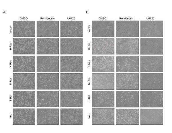
Figure 5
Romidepsin more significantly alters the morphology of RIE-1 cells than that of NIH 3T3 cells. NIH 3T3 (A) or RIE-1 (B) cells stably expressing the indicated proteins were incubated in complete growth medium supplemented with DMSO (Vehicle), 5 nM romidepsin, or 10 μM U0126 for 48 h. Representative fields of cells were photographed under 10× magnification. Data shown are representative of at least two independent experiments.
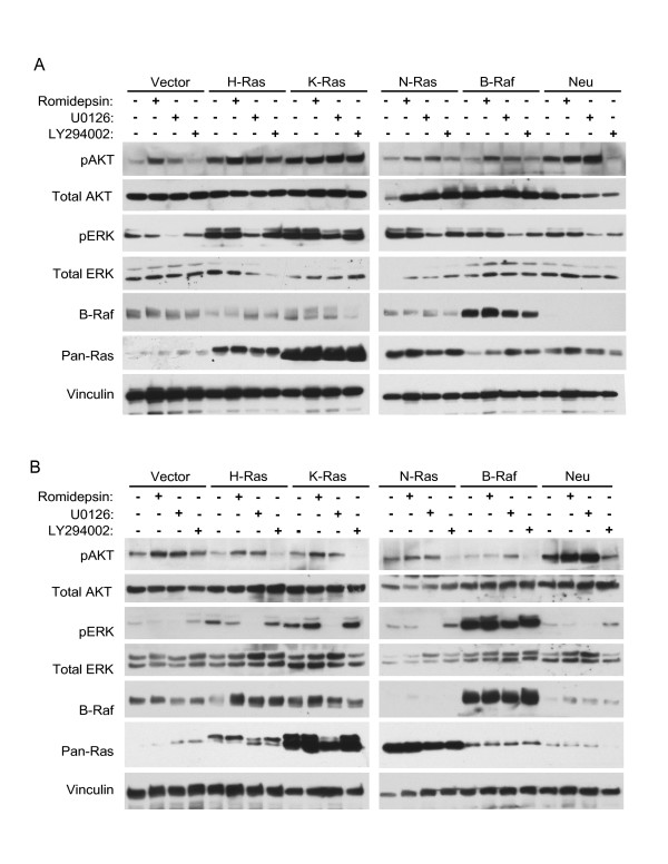
Figure 6
Romidepsin does not inhibit Ras-mediated ERK or AKT activation in NIH 3T3 and RIE-1 cells. NIH 3T3 (A) or RIE-1 (B) cells stably expressing the indicated proteins were treated with DMSO (Vehicle), 5 nM romidepsin, 10 μM U0126, or 10 μM LY294002 for 24 h. Cell lysates were analyzed by western blotting with antibodies to total ERK1/2, phospho-ERK1/2, total AKT, phospho-Akt, B-Raf, pan-Ras, and vinculin (loading control). Data shown are representative at least two independent experiments.