
Figure 1
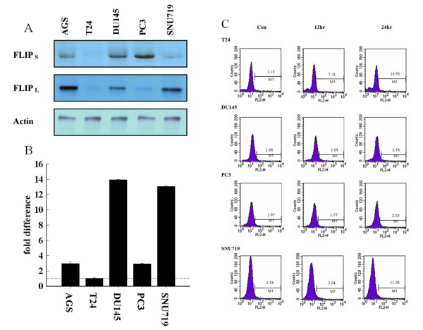
The expression levels of c-FLIPS indicate a relationship to resistance to TNF-induced apoptosis. (A) Immunoblotting analysis of c-FLIPL and c-FLIPS in TNF-α-resistant and TNF-α-sensitive cell lines with anti-FLIPL and anti-FLIPS antibodies. The molecular weights of c-FLIPL and c-FLIPS were about 55 kDa and 28 kDa, respectively. β-Actin was used as a loading control. (B) Differences in the expression levels of c-FLIPS between various cancer cell lines shown by quantitative real-time PCR and SYBR Green detection. Levels of c-FLIPS were normalized to GAPDH and the fold changes in c-FLIPS levels are shown for each cell line. Data shown are the average of two assays. PCR products were analyzed using Sequence Detection software. Error bars equal the SEM. (C) Measurement of TNF-α-induced apoptosis using the TUNEL assay (M, TUNEL positive) in different human cancer cell lines. Different patterns are shown following treatment with TNF-α (10 ng/ml) at 0, 12, and 24 h.
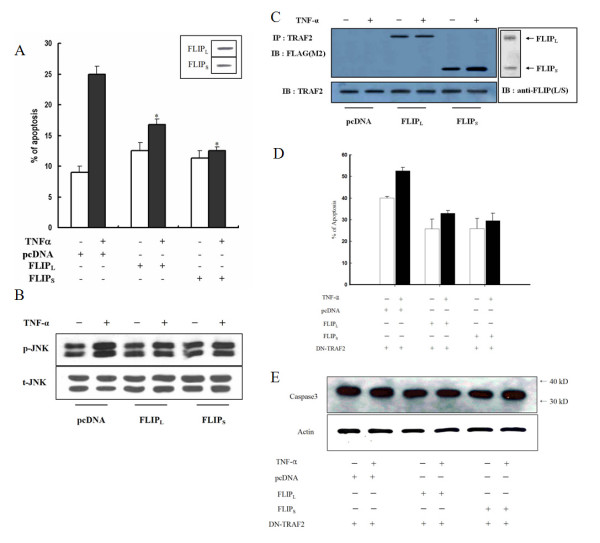
Figure 2
Expression of c-FLIPS confers stronger resistance to TNF-α-induced apoptosis than c-FLIPL, through a TRAF2-dependent mechanism. (A) Analysis of TNF-α-induced apoptosis. SNU-719 cells were transfected with the indicated constructs and treated with 10 ng/ml TNF-α for 24 h. Apoptotic cells were evaluated 48 h after transfection for each construct using the DNA fragmentation assay as described in Materials and methods. After treatment with TNF-α, the percentage of apoptotic cells transfected with pcDNA and c-FLIPL increased, but the percentage of apoptotic cells did not change in cells transfected with c-FLIPS. This experiment was performed three times independently. *P < 0.01 compared with FLIPL and FLIPS. (B) Ectopic expression of c-FLIPL by itself induced the activation of JNK in response to TNF-α, whereas c-FLIPS did not change this effect. P- and t-JNK indicate the phosphorylated JNK and total JNK levels, respectively. (C) SNU-719 cells were transfected with FLAG-tagged c-FLIPL and c-FLIPS expression vectors, incubated with TNF-α (10 ng/ml) for 12 h, and subjected to immunoprecipitation with anti-TRAF2 antibody followed by immunoblotting with anti-FLAG antibody. Immunoblotting of TRAF2 was used as a loading control. (D) To verify the role of TRAF2 in TNF-α-induced apoptosis, SNU-719 cells were cotransfected with the indicated constructs with DN-TRAF2. Apoptotic cells were evaluated 24 h after transfection for each construct using flow cytometric analysis as described in Materials and methods. (E) To confirm the results, we examined caspase 3 activity in the same conditions as described in Fig.2. D. β-Actin was used as a loading control.
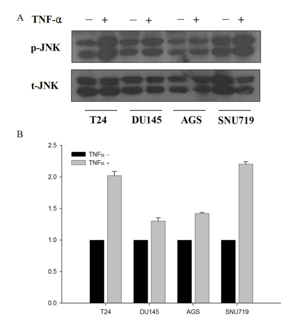
Figure 3
JNK activation in TNF-resistant cell lines. TNF-α-sensitive cell lines can activate JNK in response to TNF-α. In T24 and SNU719 cells, JNK is activated in response to TNF-α, whereas DU145 and AGS cells do not show this induction. (A) To examine the activation of JNK, protein extracts from the indicated cell lines were incubated for 6 h with TNF-α (10 ng/ml) and subjected to immunoblotting with phospho-specific and total JNK antibodies. P- and T- indicate phosphorylated proteins and total proteins, respectively. (B) Expression was quantified by densitometric scan of the blots (A). Values are the ratio of p-JNK and t-JNK and are presented relative to each control.
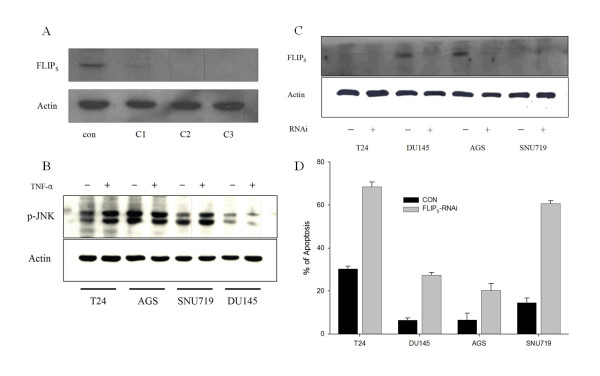
Figure 4
Knockdown of endogenous c-FLIPS in several cell lines enhances apoptosis induction by TNF-α. (A) To examine the role of c-FLIPS in TNFR1-mediated apoptosis, PC3 cells were transfected with three different siRNA oligonucleotides designed to knock down c-FLIPS. The level of c-FLIPS protein was inhibited completely by C2 and C3. In further studies, we used C3 oligonucleotide as an siRNA. (B) The indicated siRNA-transfected cells were treated with TNF-α (5 ng/ml) for 24 h and extracted for immunoblotting with phospho-specific antibodies to JNK and actin. (C) The indicated siRNA-transfected cell lines were treated with TNF-α (5 ng/ml) for 24 h before harvesting for flow cytometric analysis. This experiment was performed independently three times. (D) The indicated siRNA-transfected cell lines were treated with TNF-α (5 ng/ml) for 24 h and extracted for immunoblotting with anti-FLIPS antibody. β-Actin was used as a loading control.
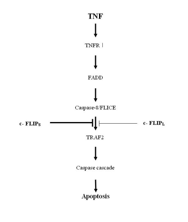
Figure 5
A working model of the role of c-FLIPS in TNFR1-mediated apoptosis. The binding of c-FLIPS to TRAF2 delays the recruitment of c-FLIPL, and c-FLIPS and c-FLIPL block TNFR1-mediated apoptosis. However, the cellular c-FLIPS level is elevated sufficiently to block apoptosis and promote cell survival.