
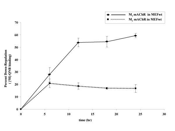
Figure 1
Time-course of agonist-promoted down-regulation of mAChRs in MEF. MEF wt cells were transfected with eGFP-M1 (◆) or HA-M2 (■) mAChR. 24 hr following transfection, cells were treated with 1 mM carbachol for the indicated time. Down-regulation was determined using [3H]-QNB binding (fmol/mg protein) in crude membranes as described in methods. The M1 mAChR displays nearly threefold the down-regulation of the M2 subtype over the same time course. Data are expressed as percent down-regulation compared to t = 0 control and are presented as mean ± standard error of the mean for three independent experiments with duplicate data points. Total mAChR expressed (fmol/mg) at t = 0 was 4000 for both M1 and M2 mAChRs.
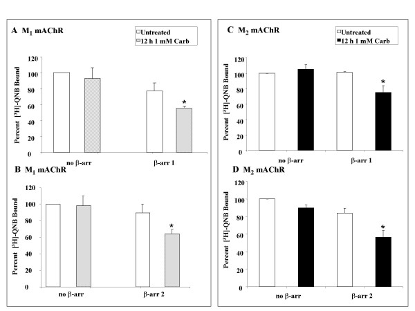
Figure 2
Rescue of mAChR down-regulation in MEF KO1/2 with β-arrestin 1 or 2. MEF KO1/2 cells were transfected with eGFP-M1 mAChR (A-B) or HA-M2 mAChR (C-D) and either FLAG-β-arrestin 1 (top) or 2 (bottom). 24 hr following transfection, cells were treated with 1 mM carbachol for 12 hr. Down-regulation was determined using [3H]-QNB binding (fmol/mg protein) in crude membranes as described in methods. No down-regulation occurs in the absence of β-arrestin and either isoform (β-arrestin 1 or 2) rescues both constitutive and agonist-promoted down-regulation. Data are expressed as percent of [3H]-QNB bound (fmol/mg total protein) compared to untreated, no β-arrestin control and are presented as mean ± standard error of the mean from three independent experiments with duplicate data points. Statistical analysis was performed using a paired t-test. *: p ≤ 0.05 versus the untreated, no β-arrestin control. Total M1 mAChR expressed (fmol/mg) at t = 0 was 400 – 900 (M1) and 1000 – 2000 (M2) for both the β-arrestin 1 and β-arrestin 2 rescue experiments.
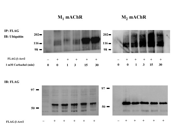
Figure 3
Agonist treatment promotes ubiquitination of β-arrestin 2 in MEF KO1/2 cells expressing the mAChRs and β-arrestin 2. MEF KO1/2 cells transfected with FLAG-β-arrestin 2 and either eGFP-M1 or HA-M2 mAChR. 24 hr following transfection, cells were treated with 1 mM carbachol for the indicated times. Top panel: cells were lysed and immunoprecipitated (IP) with anti-FLAG MAb and blotted (IB) with anti-ubiquitin-MAb. There was an increase in ubiquitination of β-arrestin 2 with increasing exposure to 1 mM carbachol. Each blot is representative of two independent experiments. Bottom panel: lysates blotted with anti-FLAG MAb to demonstrate β-arrestin 2 expression levels.
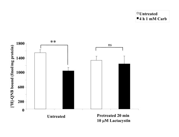
Figure 4
The proteosomal inhibitor lactacystin inhibits the β-arrestin 2 mediated agonist-promoted down-regulation of the M2 mAChR in MEFwt. MEFwt cells were transfected with HA-M2 mAChR and, after 24 hr cells were incubated for 20 min with or without 10 μM lactacystin then treated with 1 mM carbachol for 4 hr. Lactacystin inhibits the β-arrestin 2 mediated down-regulation of the M2 mAChR receptor in response to agonist. Down-regulation was determined using [3H]-QNB binding in crude membranes as described in methods. Data are expressed as [3H]-QNB bound (fmol/mg total protein) and are presented as mean ± standard deviation from three independent experiments with duplicate data points. Statistical analysis was performed using a paired t-test, ** indicates p ≤ 0.001 (compared to paired untreated control), ns indicates not significant.
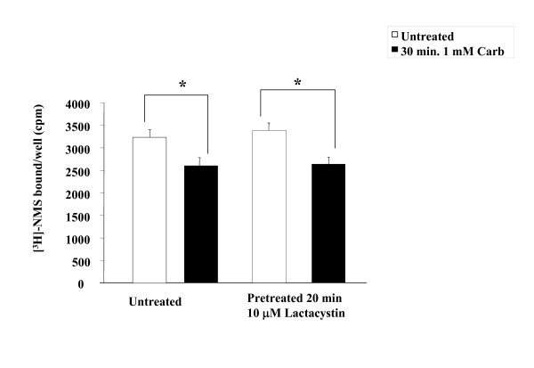
Figure 5
The proteosomal inhibitor lactacystin does not affect agonist-promoted down-regulation internalization of the M2 mAChR in MEFwt. MEFwt cells were transfected with HA-M2 mAChR and, after 24 hr cells were incubated for 20 min with or without 10 μM lactacystin then treated with 1 mM carbachol for 30 min. Lactacystin has no effect on the internalization of the M2 mAChR receptor in response to agonist. Internalization was determined using [3H]-NMS binding in whole cells as described in methods. Data are [3H]-NMS bound per well (plated at 1 × 10⁵ cells) and are presented as mean ± standard deviation from three independent experiments with duplicate data points. Statistical analysis was performed using a paired t-test, * indicates p ≤ 0.05; (compared to paired untreated control), ns indicates not significant.
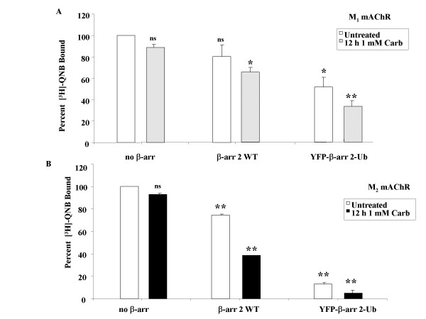
Figure 6
Expression of a chimeric ubiquitinated form of β-arrestin 2 enhances the agonist-promoted down-regulation of M1 and M2 mAChRs in MEF KO1/2. MEF KO1/2 cells were transfected with eGFP-M1 (A) or HA-M2 (B) mAChRs and either no β-arrestin, FLAG-β-arrestin 2 or YFP-β-arrestin 2-Ub. 24 hr following transfection, cells were treated with 1 mM carbachol for 12 hr. Down-regulation was determined using [3H]-QNB binding (fmol/mg protein) in crude membranes as described in methods. Expression of the constitutively ubiquitinated form of β-arrestin (β-arrestin 2-Ub) had a greater effect on down-regulation of the M2 mAChR compared to the M1 subtype. Data are expressed as percent of [3H]-QNB bound compared to the untreated control with no β-arrestin and are presented as mean ± standard deviation from two independent experiments with duplicate data points. Statistical analysis was performed using a repeated measures ANOVA with Bonferroni post test; * indicates p ≤ 0.05 and ** indicates p ≤ 0.001 (compared to untreated, no β-arrestin control), ns indicates not significant. Total mAChR expressed (fmol/mg) in the absence of β-arrestin was 1000 – 2000 for both receptor subtypes.
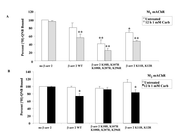
Figure 7
β-arrestin 2K18R, K107R, K108R, K207R, K296Rdifferentially affects down-regulation of M1 and M2 mAChRs in MEF KO1/2. MEF KO1/2 cells were transfected with eGFP-M1 (A) mAChR or HA-M2 (B) mAChR and either empty vector (control), FLAG-β-arrestin 2 (WT), FLAG-β-arrestin 2K18R, K107R, K108R, K207R, K296R or FLAG-β-arrestin 2K11R, K12R. 24 hr after transfection, cells were treated with 1 mM carbachol for 12 hr. Down-regulation was determined in crude membranes (fmol/mg protein) as described in methods. All β-arrestin 2 constructs were able to mediate agonist-promoted down-regulation of mAChR with the exception of the β-arrestin 2K18R, K107R, K108R, K207R, K296R mutant when co-expressed with the M2 mAChR. Data are expressed as percent of [3H]-QNB bound (compared to untreated, no β-arrestin control) and presented as mean ± standard deviation from three independent experiments with duplicate or quadruplicate data points. Statistical analysis was performed using a repeated measures ANOVA with Bonferroni post test; * indicates p ≤ 0.05 ** indicates p ≤ 0.001 (compared to untreated, no β-arrestin control), ns indicates not significant. Total M1 (300–500 fmol/mg) and M2 (1500–2500 fmol/mg) mAChR expressed in the absence of β-arrestin constructs was similar.
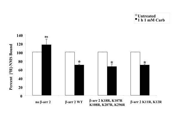
Figure 8
Agonist-promoted internalization of M2 mAChR is unaffected by β-arrestin 2 lysine mutants in MEF KO1/2. MEF KO1/2 cells were transfected with HA-M2 mAChR and either empty vector (control), FLAG-β-arrestin 2 (WT), FLAG-β-arrestin 2K18R, K107R, K108R, K207R, K296R or FLAG-β-arrestin 2K11R, K12R. 24 hr after transfection, cells were treated with 1 mM carbachol for 1 hr. Internalization was determined in whole cells (receptors/cell) as described in methods. All constructs were able to mediate agonist-promoted internalization. Data are expressed as percent of [3H]-NMS bound compared to untreated control and presented as mean ± standard deviation from three independent experiments with duplicate data points. Statistical analysis was performed using a repeated measures ANOVA with Bonferroni post test; * indicates p ≤ 0.05 (compared to untreated, no β-arrestin control), ns indicates not significant. Total receptor expressed in the presence of all four β-arrestin constructs was between 5 and 6 × 10⁵ receptor/cell.
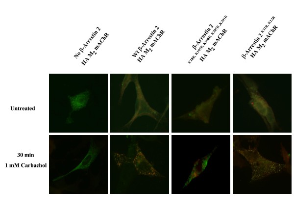
Figure 9
β-arrestin 2K18R, K107R, K108R, K207R, K296Rdemonstrates impaired agonist-promoted co-localiztion with M2 mAChR in MEF KO1/2. MEF KO1/2 cells were transfected with HA-M2 mAChRs (green) and either wild-type FLAG-β-arrestin 2, FLAG-β-arrestin 2K18R, K107R, K108R, K207R, K296R or FLAG-β-arrestin 2K11R, K12R (red). 24 hr after transfection, cells were treated with 1 mM carbachol for 30 min. Cells were fixed, probed, and imaged as described in methods. Wild-type and β-arrestin 2K11R, K12R demonstrate co-localization with the M2 mAChR which is significantly diminished with β-arrestin 2K18R, K107R, K108R, K207R, K296R. Image shown is representative of two independent experiments.
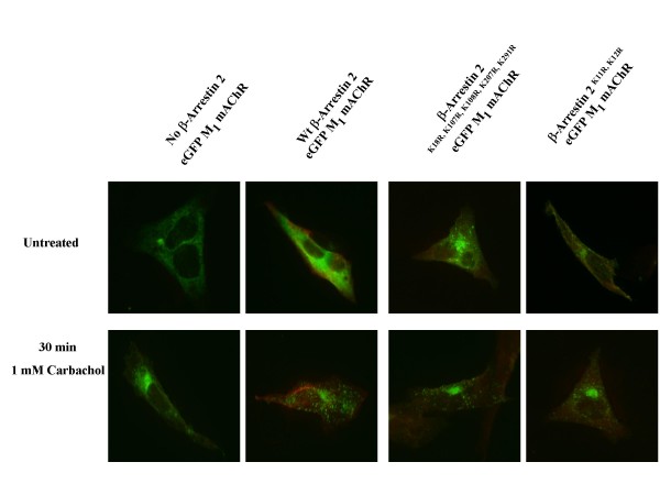
Figure 10
β-arrestin 2K18R, K107R, K108R, K207R, K296Rdemonstrates no co-localization with M1 mAChR in MEF KO1/2. MEF KO1/2 cells were transfected with eGFP-M1 (A) mAChRs (green) and either wild-type FLAG-β-arrestin 2, FLAG-β-arrestin 2K18R, K107R, K108R, K207R, K296R or FLAG-β-arrestin 2K11R, K12R (red). 24 hr after transfection, cells were treated with 1 mM carbachol for 30 min. Cells were fixed, probed, and imaged as described in methods. None of the β-arrestin constructs demonstrate co-localization with the M1 mAChR. Image shown is representative of two independent experiments.
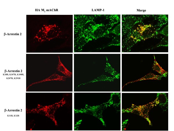
Figure 11
β-arrestin 2K18R, K107R, K108R, K207R, K296Rdisrupts the co-localization of M2 mAChR and LAMP-1 in MEF KO1/2. MEF KO1/2 cells were transfected with HA-M2 mAChR and either wild-type FLAG-β-arrestin 2, FLAG-β-arrestin 2K18R, K107R, K108R, K207R, K296R or FLAG-β-arrestin 2K11R, K12R. 24 hr after transfection, cells were treated with 1 mM carbachol for 6 hr. Cells were fixed, probed, and imaged as described in methods. β-arrestin 2K18R, K107R, K108R, K207R, K296R disrupts the co-localization of M2 mAChR and LAMP-1 compared to wild-type and β-arrestin 2K11R, K12R. Image shown is representative of three independent experiments.