
Figure 1
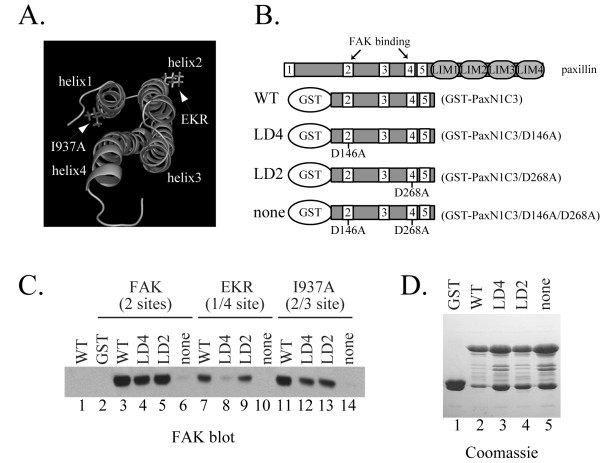
In vitro binding of paxillin and FAK mutants. A) The I937A and EKR mutations are indicated on the structure of the FAT domain. The four α-helices are labeled. The paxillin binding sites are at the interface of α-helices 1/4 and the interface of α-helices 2/3. B) Schematic representation of paxillin and paxillin GST-fusion proteins used for the binding studies. White boxes denote the LD motifs. The proteins are designated WT, LD4, LD2 and none to reflect functional LD motifs in the constructs. The previously published designation of each construct is in parentheses. C) CE cells transfected with empty RCAS vector (lane 1), wild type FAK (lanes 2–6), EKR (lanes 7–10), or I937A (lanes 11–14) were lysed and 1 mg of lysate was used for binding assays. The functional paxillin binding site in each of the constructs is indicated in parentheses. Lysates were incubated with GST alone (lane 2), the WT (lanes 3, 7, 11), the LD4 (lanes 4, 8, 12), the LD2 fusion proteins (lanes 5, 9, 13), or with the protein lacking both LD2 and LD4 sites (none; lanes 6, 10, 14). The protein complexes were washed and bound FAK detected by Western blotting. D) GST-fusion proteins used in C were analyzed in parallel by SDS-PAGE and Coomassie staining to ensure equal loading.
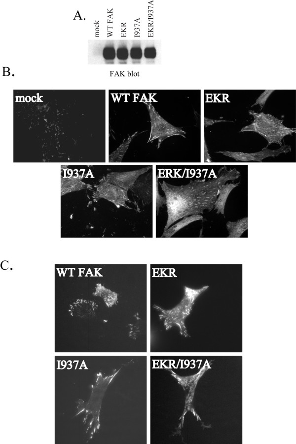
Figure 2
Subcellular localization of FAK mutants. A) CE cells transfected with empty RCAS vector (mock) or RCAS constructs encoding wild type FAK or FAK mutants were lysed and 25 μg of lysate was blotted for FAK expression with BC4 antibody. B) CE cells transfected with empty RCAS vector (mock) or RCAS encoding wild type FAK or FAK mutants were plated on fibronectin-coated coverslips overnight, then fixed and used for immunofluorescent imaging using the FAK BC4 antibody. C) CE cells transfected with GFP-fusion constructs of wild type FAK or mutants were visualized in live cells by TIRF microscopy.
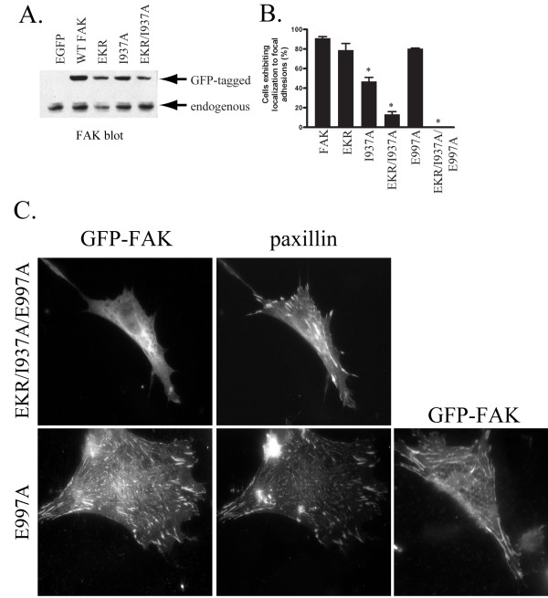
Figure 3
Quantification of focal adhesion localization. A) CHO-K1 cells were transfected with the empty EGFP vector or GFP N-terminally tagged versions of wild type or FAK mutants. Stably expressing populations were selected and lysed. Twenty-five μg of lysate was immunoblotted to examine FAK expression. Endogenous FAK and GFP-FAK are indicated. B) CHO-K1 cells stably expressing the EGFP vector alone, or GFP-FAK fusion proteins were plated on fibronectin coated coverslips overnight, then fixed and used for immunofluorescent staining with a GFP antibody. The cells were counterstained with a paxillin antibody to locate focal adhesions. GFP-positive cells were scored for GFP localization at focal adhesions. The data was analyzed using a one-way analysis of variance and the Dunnett post test (* denotes p < 0.01). C) CE cells transfected with GFP-FAK fusion proteins were plated on fibronectin-coated coverslips overnight, then fixed and used for immunofluorescent staining with a GFP antibody. The cells were also stained with a paxillin antibody to facilitate identification of focal adhesions. A second example of GFP-E997A localized to focal adhesion is also shown (lower right panel).
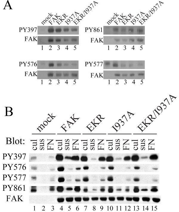
Figure 4
Identification of defects in FAK phosphorylation. A) CE cells were transfected with the RCAS empty vector (mock) or RCAS encoding wild type FAK or FAK mutants. Subconfluent cells growing in culture were lysed and immunoblotted with the indicated phospho-specific antibodies. In parallel lysates were blotted for FAK expression as a loading control. B) Phosphorylation of FAK variants was examined in cells growing in culture (cul)(lanes 1, 4, 7, 10, 13), after incubation in suspension for 30 minutes (sus)(lanes 2, 5, 8, 11, 14), and after plating on fibronectin for 45 minutes (FN)(lanes 3, 6, 9, 12, 15). Twenty-five μg of lysate was analyzed by immunoblotting using phospho-specific antibodies. Lysates were also blotted for FAK as a loading control.
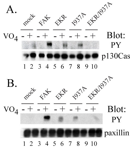
Figure 5
Phosphorylation of FAK substrates in vivo. CE cells transfected with empty RCAS vector (mock) or RCAS encoding wild type FAK or FAK mutants were left untreated (-) or treated with vanadate overnight prior to lysis (+). A) p130CAS was immunoprecipitated from 1 mg lysate and the immunoprecipitated protein was immunoblotted for phosphotyrosine (upper panel) and p130CAS (bottom panel). B) Paxillin was immunoprecipitated from 1 mg lysate and the immunoprecipitated protein was immunoblotted for phosphotyrosine (top panel) and paxillin (bottom panel).