
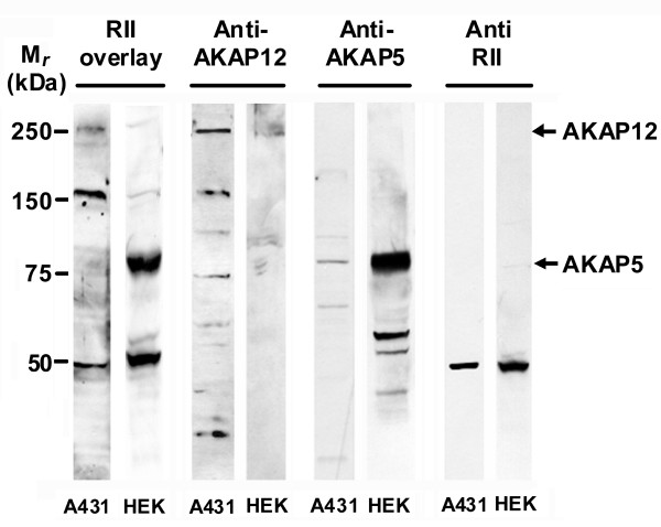
Figure 1
Overlay analysis of A kinase-anchoring proteins in A431 and HEK cells. Cell lysates (50 μg of protein/lane) of A431 or HEK cells were subjected to SDS-polyacrylamide gel electrophoresis, the resolved proteins transferred to nitrocellulose blots, and the blots subjected to renaturation and stained with A-kinase RII subunit for 2 h at room temperature. After washing, the presence of AKAPs was detected using an goat anti-RII antibody. The resultant blots also were stained with antibodies specific for AKAP12 or AKAP5.
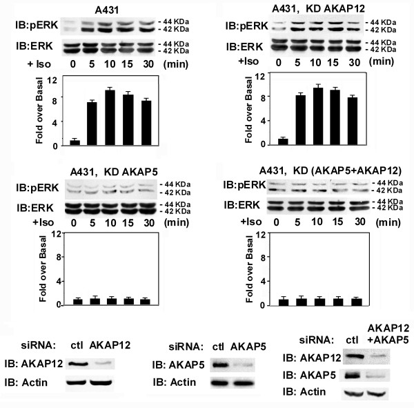
Figure 2
Activation of the mitogen-activated protein kinase cascade to Erk1,2 in human epidermoid A431 cells in response to isoproterenol stimulation. Wild-type 431 or A431 cells treated with siRNAs targeting either AKAP12 or AKAP5, or both AKAPs, were stimulated by isoproterenol (10 μM) for different intervals. Cell lysates were subjected to SDS-polyacrylamide gel electrophoresis, the resolved proteins transferred to nitrocellulose. The blots were stained by antibody against activation-specific phospho-Erk1,2, against pan-Erk, or against AKAP12 and AKAP5.
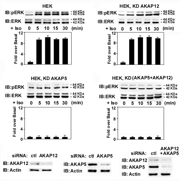
Figure 3
Activation of the mitogen-activated protein kinase cascade to Erk1,2 in human embryonic kidney 293 cells in response to isoproterenol stimulation. Wild-type HEK293 cells or HEK293 cells treated with siRNAs targeting either AKAP12 or AKAP5, or both AKAPs, were stimulated by isoproterenol (10 μM) for different intervals. Cell lysates were subjected to SDS-polyacrylamide gel electrophoresis, the resolved proteins transferred to nitrocellulose. The blots were stained by antibody against activation-specific phospho-Erk1,2, against pan-Erk, or against AKAP12 and AKAP5.
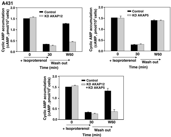
Figure 4
Desensitization and Resensitization of β2AR in wild-type A431 cells and cells treated to knock-down AKAP12 or AKAP5. A, cyclic AMP accumulation of β2AR-mediated cyclic AMP accumulation in A431 cells that were desensitized by 30 min treatment with Iso and then washed free of agonist for 60 min (W60) to permit resensitization was examined. Agonist-stimulated (Iso,10 μM) cyclic AMP accumulation was measured in untreated cells, in cells following a 30-min prior stimulation with Iso, and in cells treated with Iso for 30 min, washed free of agonist, incubated for 60 min and then assayed (W60). A431 cells were pretreated either with a control, scrambled sequence siRNA (Control) or with siRNA targeting AKAP12/AKAP5 to knock down (KD) the expression of endogenous AKAP12 or AKAP5. Cells were challenged with a β2-adrenergic agonist (Iso, 10 μM) for 30 min to provoke agonist-induced desensitization and internalization of β2AR. The recovery from agonist-induced desensitization, termed resensitization, was measured in cells after a 30-min challenge with Iso, and in Iso-treated cells that were washed free of agonist for 60 min (W60). Isoproterenol-stimulated cyclic AMP accumulation was measured in these cells and is reported as picomol of cyclic AMP accumulated per 105 cells. The results, displayed as mean ± S.E., are derived from at least three separate experiments performed with as many separate cultures of A431 cells. *, p ≤ 0.01 for the difference from the amount of cyclic AMP accumulation measured in the control cells at 60 min following a washout of the agonist (W60).
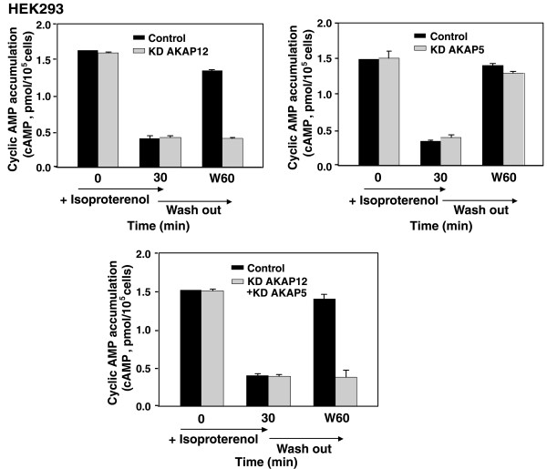
Figure 5
Desensitization and Resensitization of β2AR in wild-type HEK293 cells and cells in which AKAP12 or AKAP5 were knocked-down. A, cyclic β2AR-mediated cyclic AMP accumulation in HEK cells that were desensitized by 30 min treatment with Iso and then washed free of agonist for 60 min (W60) to permit resensitization was examined. Agonist-stimulated (Iso,10 μM) cyclic AMP accumulation was measured in untreated cells, in cells following a 30-min prior stimulation with Iso, and in cells treated with Iso for 30 min, washed free of agonist, incubated for 60 min and then assayed (W60). HEK cells were pretreated with either a control, scrambled sequence siRNA (Control) or with siRNA to AKAP12 or AKAP5 to knock down (KD) the expression of endogenous AKAP12 or AKAP5. Cells were challenged with a β2-adrenergic agonist (Iso,10 μM) for 30 min to provoke agonist-induced desensitization and internalization of β2AR. The recovery from agonist-induced desensitization, termed resensitization, was measured in cells after a 30-min challenge with Iso, and in Iso-treated cells that were washed free of agonist for 60 min (W60). Isoproterenol-stimulated cyclic AMP accumulation was measured in these cells and is reported as picomol of cyclic AMP accumulated per 105 cells. The results, displayed as mean ± S.E., are derived from at least three separate experiments performed with as many separate cultures of A431 cells. *, p ≤ 0.01 for the difference from the amount of cyclic AMP accumulation measured in the control cells at 60 min following a washout of the agonist (W60).
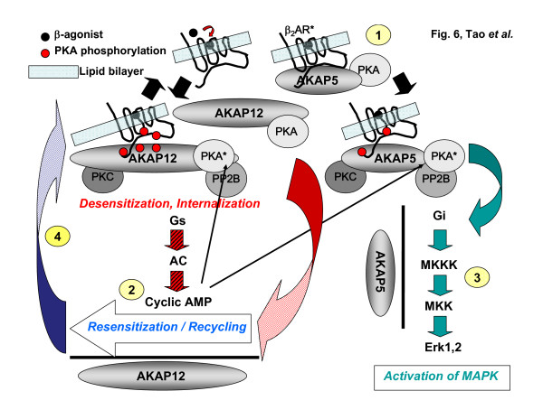
Figure 6
Schematic of GPCR-associated AKAP79 and AKAP250: regulation of downstream signaling by beta2-adrenergic receptors. The AKAPs provide multivalent docking sites for PKA, PKC, and PP2B as well as other protein kinases, phosphoprotein phosphatases, and adaptor molecules (not shown). Binding of the beta-adrenergic agonist leads to activation of the receptor (β2AR*, #1) and activation of Gs > adenylylcyclase activation > cyclic AMP accumulation > PKA activation (#2). The association of AKAP250 with the receptor is markedly enhanced by PKA phosphorylation of the receptor and scaffold, whereas AKAP79 association with the receptor is constitutive. AKAP79 mediates downstream signal switching from Gs to Gi-mediated activation of the mitogen-activated protein kinase cascade leading to Erk1,2 (#3). AKAP250 mediates the resensitization and recycling of the desensitized and internalized receptor (#4). Through docking of critical enzymes, the AKAP provide high spatial resolution and localization of downstream signaling. See the text for more details.