
Figure 1
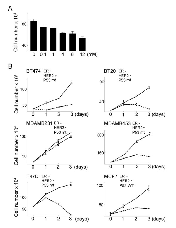
Metformin inhibits proliferation of cultured breast cancer cells. A. MCF7 cells were treated with metformin at the indicated concentrations for one day and then cell number was determined using a hemocytometer. The mean cell number from 3 independent cultures is shown. Error bars represent standard deviation. Using a t test, all treatments were significantly different from the control with P values of less than 0.01. B. Six different breast cancer cell lines were treated with 8 mM metformin. At the indicated times viable cell number was determined using a hemocytometer. The reported status of estrogen receptor (ER) expression, HER2 amplification, and p53 expression (Wild-type, WT, or mutant, mt) are indicated for each cell line. HER2+ indicates HER2 amplification while HER2- indicates normal HER2 expression. The data points represent the mean cell number from 3 independent cultures and error bars represent standard deviation.
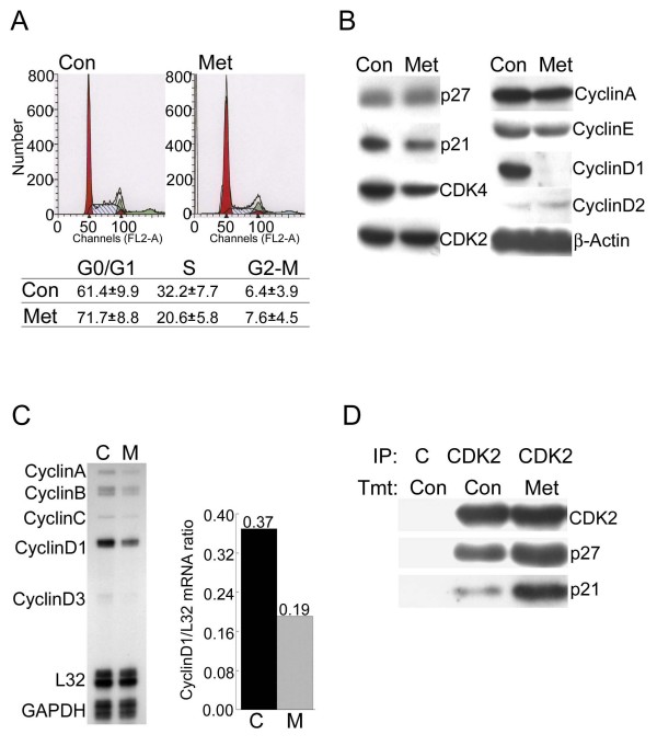
Figure 2
Effect of metformin on cell cycle progression and cell cycle regulatory proteins in MCF7 cells. A. Untreated cells (Con) or cells treated with 8 mM metformin for 1.5 days (Met) were stained with propidium iodide and then analyzed by flow cytometry to estimate the number of cells in each phase of the cell cycle. The experiment was repeated 3 times and the mean and standard error for each cell phase is indicated in the table. B. Equal amounts of protein from untreated cells (Con) or cells treated with metformin for 1.5 days (Met) were analyzed by western blotting using antibodies that recognize the indicated cell cycle regulatory proteins. β-Actin was detected as a loading control C. Total RNA was isolated from untreated cells (C) or cells treated with metformin (8 mM) for 1.5 days (M) and RNase protection assays were performed to detect the mRNAs encoding the indicated cyclins (left panel). Cyclin D1 mRNA levels were normalized using L32 mRNA levels (right panel). The mean of 3 independent experiments is shown and error bars indicate standard deviation. In a t test the value for the metformin treated cells was significantly different from the control with a P value of 0.006. D. MCF7 cells were treated with (Met) or without (Con) metformin (8 mM) and then extracts were prepared for immunoprecipitation (IP) using either a control antibody (C) or an anti-CDK2 antibody (CDK2). The immunoprecipitated proteins were analyzed by western blotting using antibodies that recognize CDK2, p27Kip1 or p21Cip1 as indicated on the right.
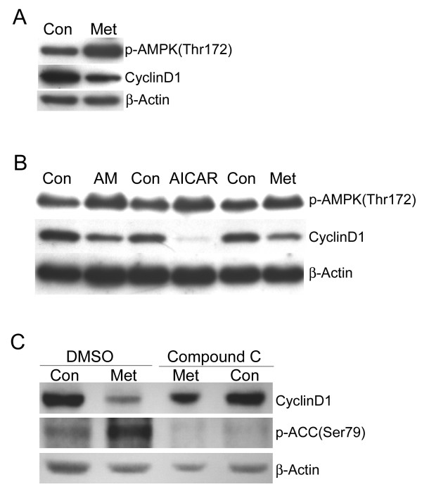
Figure 3
Downregulation of cyclin D1 corresponds to activation of AMP-activated protein kinase. A. MCF7 cells were treated with (Met) or without (Con) metformin for 1.5 days and then western blotting was performed to detect active phospho-AMPK, cyclin D1, and β-actin. B. MCF7 cells were treated with antimycin A (AM, 1 μM), AICAR (4 mM), or metformin (8 mM) for 1.5 days. Control cells (Con) were treated with the appropriate vehicle for each reagent. Western blotting was performed to detect active phospho-AMPK, cyclin D1, and β-actin. C. MCF7 cells were pretreated with DMSO (vehicle) or the AMPK-specific inhibitor compound C (20 μM) for 1 day and then treated with (Met) or without (Con) metformin for 1.5 days. Western blotting was performed to detect cyclin D1 or the phosphorylated form of the AMPK substrate ACC. β-actin was used as a loading control.
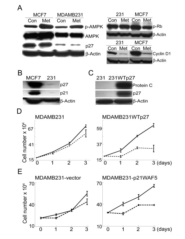
Figure 4
Overexpression of p27Kip1 in the metformin-resistant MDA-MB-231 cell line leads to metformin sensitivity. A. (Left panel) MCF7 and MDA-MB-231 cells were treated with (Met) and without (Con) metformin (8 mM) for 2 days. Western blotting was used to detect phospho-AMPK, total AMPK, p27Kip1, and β-actin. (Right panel) Same as A except that cell extracts were used to detect phospho-Rb (Serine 795), cyclin D1 or β-actin. B. Extracts from untreated MCF7 and MDA-MB-231 cells were used for western blotting of p27Kip1, p21Cip1, and β-actin. C. MDA-MB-231 cells were stably transfected with a construct encoding p27Kip1 that was tagged with a protein C epitope to derive the cell line MDA-MB-231WTp27. Extracts from the parental cell line (231) and the stably transfected cell line (231WTp27) were used for western blotting to detect the epitope-tagged p27Kip1 (anti-Protein C epitope), p27Kip1 (p27), and β-actin. D. MDA-MB-231 cells or MDA-MB-231WTp27 cells were treated with (dashed lines) or without (solid lines) metformin (8 mM) for up to 3 days. At the indicated time points cell number was determined using a hemocytometer. Each data point represents the mean cell number from 3 independent cultures and error bars represent standard deviation. E. MDA-MB-231 cells were transiently transfected with a construct encoding p21Cip1 or empty vector. They were then treated with metformin for the indicated time and cell number was determined as describe in D.
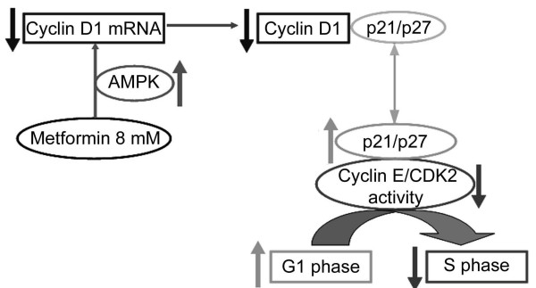
Figure 5
Proposed model for the mechanism by which metformin mediates cell cycle arrest. Metformin treatment leads to activation of AMPK which leads to loss of cyclin D1 mRNA and downregulation of cyclin D1 protein. The reduction in cyclin D1 results in the release of sequestered cell cycle inhibitors p27Kip1 and p21Cip1. The released CDK inhibitors bind to and inhibit cyclin E/CDK2, thus preventing cell cycle progression from G1 to S phase. In metformin-resistant MDA-MB-231 cells, levels of the CDK inhibitors are insufficient to block CDK2 even though AMPK is active and cyclin D1 is downregulated.