
Figure 1
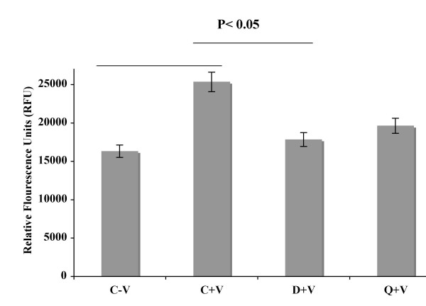
Dopamine inhibits VEGF induced in vitro permeability in HUVEC. Permeability is represented by relative fluorescence units as measured by a flux of FITC-Dextran across monolayer of HUVEC cultures in collagen-coated transwells. In the figure C-V is the control, HUVEC without any VPF/VEGF or dopamine treatment, C+V is the HUVEC treated with only VPF/VEGF (10 ng/ml) for 90 min, D+V is the HUVEC pre-treated with 10 μM dopamine for 15 min and then treated with VPF/VEGF (10 ng/ml) for 90 min and Q+V is the HUVEC pretreated with 10 μM quinpirole for 15 min and then treated with VPF/VEGF (10 ng/ml) for 90 min. Experiments were repeated three times in triplicate.
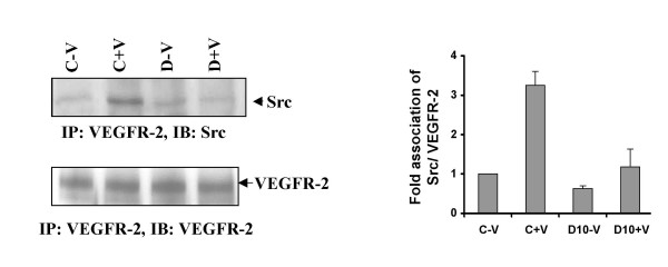
Figure 2
Dopamine treatment alters VEGF/VPF induced VEGR-2 and c-Src association in HUVEC. HUVEC extracts were immunoprecipitated with VEGFR-2 antibody and immunoblotted with Src and VEGFR-2 antibody. In the figure C-V is the control, HUVEC without any VPF/VEGF or dopamine treatment, C+V is the HUVEC treated with only VPF/VEGF (10 ng/ml) for 15 min, D-V is the HUVEC treated with 10 μM dopamine for 15 min and D+V is the HUVEC pretreated with 10 μM dopamine for 15 min and then treated with VPF/VEGF (10 ng/ml) for 15 min. Q-V is the HUVEC treated with 10 μM quinpirole for 15 min and Q+V is the HUVEC pretreated with 10 μM quinpirole for 15 min and then treated with VPF/VEGF (10 ng/ml) for 15 min. The figures are representative of three separate experiments with similar results.
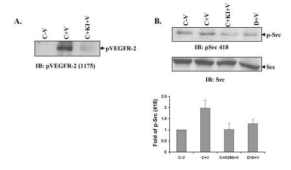
Figure 3
VPF/VEGF induced VEGFR-2 phosphorylation is required for Src phosphorylation. (A) Kinase inhibitor (250 nM) inhibits VPF/VEGF induced VEGFR-2 Y1175 phosphorylatin. In the figure C-V is control, HUVEC without any VPF/VEGF or dopamine treatment, C+V is the HUVEC treated with only VEGF (10 ng/ml) for 15 min, C+KI+V is the HUVEC pretreated with kinase inhibitor (KINASE IV) for 30 min and treated with VPF/VEGF (10 ng/ml) for another 15 min (B) Pre-treatment of HUVEC with kinase inhibitor inhibits VEGF induced Src Y418 phosphorylation. In the figure C-V is control, HUVEC without any VPF/VEGF or dopamine treatment, C+V is the HUVEC treated with only VPF/VEGF (10 ng/ml) for 15 min, C+KI+V is the HUVEC pre-treated with kinase inhibitor for 30 min and treated with VPF/VEGF (10 ng/ml) for another 15 min and D+V is the HUVEC pretreated with 10 μM dopamine for 15 min and then treated with VPF/VEGF (10 ng/ml) for 15 min. The figures are representative of three separate experiments with similar results.
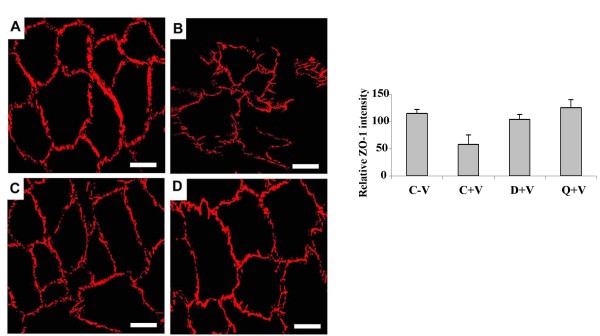
Figure 4
Confocal microscopy images of tight junctions in HUVEC. (A) normal ZO-1 in untreated control HUVEC (B) disrupted ZO-1 in VPF/VEGF (10 ng/ml) treated HUVEC (C) dopamine (10 μM) + VPF/VEGF (10 ng/ml) treated HUVEC shows that pre-treatment with dopamine inhibits the ZO-1 disrupting property of VPF/VEGF (D) quinpirole (10 μM) + VPF/VEGF (10 ng/ml) treated HUVEC shows that pre-treatment with quinpirole inhibits the ZO-1 disrupting property of VPF/VEGF. The figures are representative of three separate experiments with similar results. Average intensity of ZO-1 staining for each cell was measured using metamorph software (version 7, Molecular Devices Inc., Toronto, Canada). Scale bar, 20 μm.
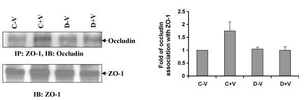
Figure 5
Effect of dopamine on ZO-1 and occludin association. Immunoprecipitation experiment showed ZO-1 and occludin were constitutively associated with each other. In the figure C-V is the control, HUVEC without any VPF/VEGF or dopamine treatment, C+V is the HUVEC treated with only VPF/VEGF (10 ng/ml) for 5 min, D-V is the HUVEC treated with 10 μM dopamine for 15 min and D+V is the HUVEC pretreated with 10 μM dopamine for 15 min and then treated with VPF/VEGF (10 ng/ml) for 5 min. The figures are representative of three separate experiments with similar results.
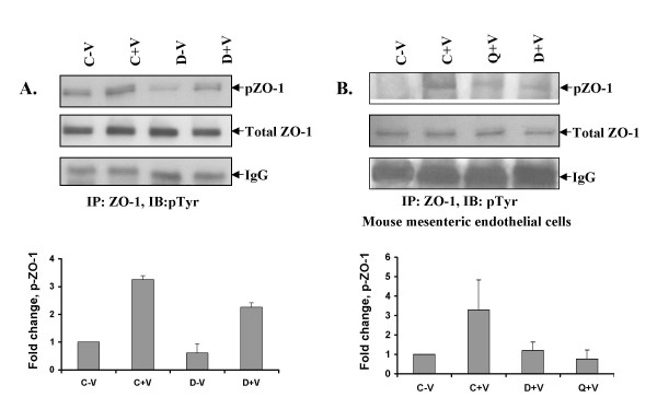
Figure 6
Effect of dopamine on VPF/VEGF-induced phosphorylation of ZO-1 in vitro and in vivo. (A) Decreased VPF/VEGF-induced phosphorylation of ZO-1 in HUVEC incubated with dopamine (10 μM) for 15 min before VPF/VEGF (10 ng/ml) treatment. In the figure C-V is control, HUVEC without any VPF/VEGF or dopamine treatment, C+V is the HUVEC treated with only VPF/VEGF (10 ng/ml) for 5 min, D-V is the HUVEC treated with 10 μM dopamine for 15 min and D+V is the HUVEC pretreated with 10 μM dopamine for 15 min and then treated with VPF/VEGF (10 ng/ml) for 5 min. The figures are representative of three separate experiments with similar results. (B) Increased VPF/VEGF-induced phosphorylation of ZO-1 in mesenteric vascular endothelium (MVE) of mice treated with 500 ng of VPF/VEGF [C+V] and decreased VPF/VEGF-induced phosphorylation of ZO-1 in MVE of mice treated with either quinpirole (10 mg/kg i.p.) [Q+V] or dopamine (50 mg/kg i.p.) [D+V] 10 min before VPF/VEGF (500 ng) treatment were detected. C-V is the untreated control.
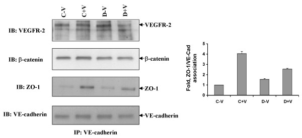
Figure 7
ZO-1, β-catenin, VE-Cadherin and VEGFR-2 are in the same immunocomplex. Immunoprecipitation with VE-cadherin antibody showed constitutive association between VE-cadherin, VEGFR-2, β-catenin and ZO-1. In the figure C-V is control, HUVEC without any VPF/VEGF or dopamine treatment, C+V is the HUVEC treated with only VPF/VEGF (10 ng/ml) for 5 min, D-V is the HUVEC treated with 10 μM dopamine for 15 min and D+V is the HUVEC pretreated with 10 μM dopamine for 15 min and then treated with VPF/VEGF (10 ng/ml) for 5 min. The figures are representative of three separate experiments with similar results.
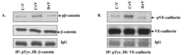
Figure 8
Dopamine pre-treatment inhibits VPF/VEGF-induced phosphorylation of β-catenin and VE-cadherin. HUVEC were pretreated with dopamine (10 μM) 15 min before VPF/VEGF (10 ng/ml) treatment. In the figure C-V is control, HUVEC without any VPF/VEGF or dopamine treatment, C+V is the HUVEC treated with only VPF/VEGF (10 ng/ml) for 5 min and D+V is the HUVEC pretreated with 10 μM dopamine for 15 min and then treated with VPF/VEGF (10 ng/ml) for 30 min. The cell extracts were immunoprecipitated (IP) with anti-phosphotyrosine antibodies and immunoblotted (IB) with either (A) anti-β-catenin or (B) anti-VE-cadherin antibodies. The figures are representative of four separate experiments with similar results.