
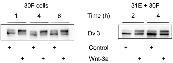
Figure 1
Dvl-3 as a marker of Wnt pathway activation. Line 30F (left panel) or co-cultures of Lines 31E + 30F (right panel) stimulated with ectopic Wnt-3a respond with hyperphosphorylation of Dvl-3. Following treatment with Wnt-3a for the indicated times, cells were lysed with Triton X-100 Lysis Buffer and cell extracts were subjected to Western blot analysis. Hyperphosphorylation of Dvl-3 is evident by a consistent relative increase in the intensity of the upper band of the doublet. Alkaline phosphatase treatment caused the loss of the upper band (data not shown).
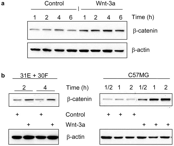
Figure 2
Accumulation of β-catenin in the cytosol upon stimulation with Wnt-3a. Line 30F cells (a), and co-cultures of Lines 31E + 30F or C57MG cells (b) were stimulated with purified Wnt-3a or mock-purified conditioned medium (Control) for the indicated times. Free cytoplasmic β-catenin was obtained by hypotonic lysis using Dounce homogenization followed by immunoblot analysis with a broadly reactive polyclonal, anti-β-catenin antibody. Increased "free" cytosolic β-catenin is evident even after half an hour of Wnt-3a treatment of C57MG cells (b), and is sustained for up to the 6 hour time point examined with 30F cells (a). Immunodetection of β-actin served as a loading control.
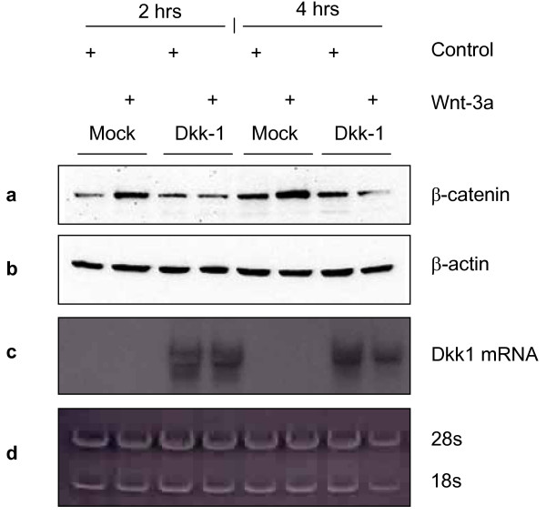
Figure 4
Inhibition of Wnt-3a pathway activation by Dkk-1. L cells were transiently transfected with an empty vector (Mock) or with a plasmid encoding Dkk-1 and were either control-treated or purified Wnt-3a-treated for 2 and 4 hours. (a) At both time points cells transfected with the Dkk-1 plasmid fail to induce accumulation of cytoplasmic β-catenin upon Wnt-3a treatment, as detected by Western Blot. In contrast, Mock transfected cells show an elevation in the level of β-catenin upon Wnt-3a treatment. (b) The detection of β-actin served as a loading control. (c) Parallel plates were treated as above and lysates were collected for extraction of total RNA. Dkk-1 mRNA is shown by Northern Blot in the cells transfected with Dkk-1. (d) 18S and 28S rRNA bands are loading controls obtained by acridine orange staining of the gel used for the Northern Blot prior to blotting.
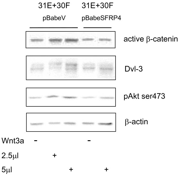
Figure 5
SFRP-4 interferes with Wnt-3a signalling in Lines 31E + 30F co-cultures. pBabeSFRP-4-infected, puromycin-selected, 31E cells in co-culture with 30F cells fail to show Wnt and Akt signalling activation by Wnt-3a. Treatment with Wnt-3a induced a hyperphosphorylation of Dvl-3, an increase in Akt phosphorylation (Ser473), and raised the levels of "active" β-catenin in the absence (pBabeV, vector), but not in the presence of the SFRP-4 cells.
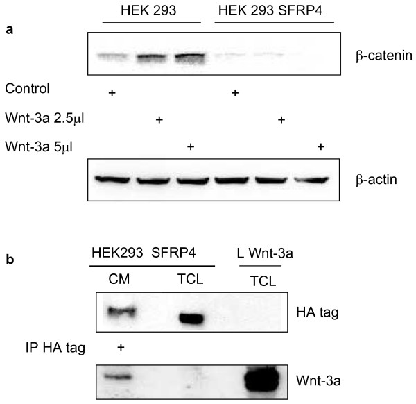
Figure 8
SFRP-4 binds to Wnt-3a, and thereby prevents activation of the β-catenin pathway. (a) HEK293 and HEK293 transfected with SFRP-4(HA tagged) were either control-treated or purified Wnt-3a-treated for 4 hours. Cytosolic lysates were analysed by Western Blot for the levels of β-catenin. Pathway activation is indicated by the increase in cytosolic β-catenin upon Wnt-3a treatment seen only in the parental HEK293 cells whereas in the SFPR-4-expressing cells this effect was abolished. (b) conditioned medium supernatant (CM) prepared from the HEK 293 cells expressing SFRP-4(HA tagged) after treatment with 5 μl Wnt-3a for 4 hours in the above experiment, was immunoprecipitated with an anti-HA-tag antibody, electrophoresed, Western blotted and, firstly, detected with an anti-HA-tag antibody. The first lane shows the precipitated SFRP-4 containing the HA tag from the CM. The middle lane shows the presence of the same protein in the total cell lysate (TCL) of the same cells as a control. The membrane was then re-probed with affinity-purified anti-Wnt-3a antibody, showing binding of Wnt-3a to the immunoprecipitated SFRP-4(HA tagged) from the conditioned medium supernatant (first lane), but not in the total cell lysate (middle lane). In addition, a sample of TCL from L cells expressing Wnt-3a (Fig. 8b, last lane), which had not been subjected to immunoprecipitation, served as a control demonstrating the detection of Wnt-3a protein.
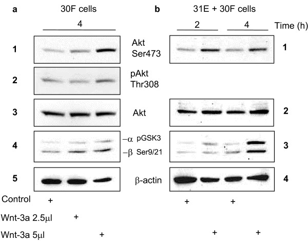
Figure 3
Wnt signalling induces phosphorylation and activation of Akt/PKB. (a) Upon stimulation with 5 μl/ml of purified Wnt-3a for 4 hours, Line 30F cells show an elevation of phoshorylated Akt in both Ser473 (panel 1) and Thr308 (panel 2) which coincides with phosphorylation of GSK3β at Ser9/21 (panel 4), reflecting its inactivation by Akt. Total Akt was used as a loading control by stripping the membrane and reprobing with an anti-Akt antibody (panel 3). β-actin was used as a loading control for pGSK-3β (panel 5). (b) Treatment of 31E + 30F co-cultures with purified Wnt-3a for 2 and 4 hours also resulted in phosphorylation of Akt at Ser473 (panel 1) and of GSK-3α and -3β at Ser9/21 (panel 3).
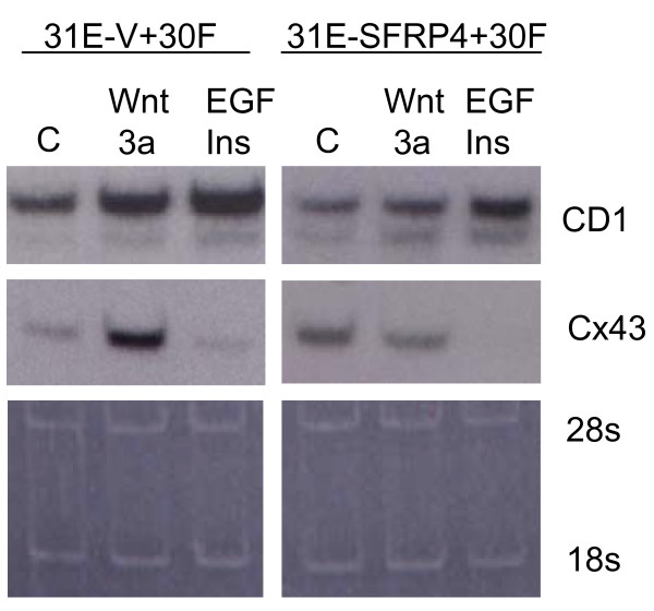
Figure 6
Cyclin D1 and connexin-43 mRNA expression in Lines 31E + 30F co-cultures after Wnt-3a stimulation. Northern blots were employed to show the expression of cyclin D1 and connexin-43 polyA+ RNAs after stimulation of the cultures for 12 hours with Wnt-3a or insulin + epidermal growth factor (EGF). Co-cultures, made with either pBabeVector or pBabeSFRP-4-infected Line 31E cells together with Line 30F cells, were maintained in 3% serum-containing medium and compared in their responses to Wnt-3a. With Line 31E-Vector cells (31E-V) in co-culture, either Wnt-3a alone or insulin + EGF (Ins+EGF) treatments elicit stimulation of cyclin D1 (CD1) mRNA expression, whereas connexin-43 (Cx43) expression responds only to Wnt-3a, and is strongly inhibited by EGF+insulin. A response to Wnt-3a was not seen; however, when co-cultures were made using Line 31E cells expressing pBabeSFRP-4 (31E-SFRP4), up-regulation of connexin-43 but not cyclin D1 was prevented. However, cyclin D1 mRNA up-regulation is reduced compared to the co-cultures containing Line 31E-Vector cells. Residual ribosomal RNA (loading control) was stained with acridine orange.
Table 1
Electrical resistance across monolayers of cultured Line 31E mammary epithelial cells
| Cell Type | Treatment | Transmonolayer resistance* (Ohms × cm 2 ) |
| 31E-pBabeVector | w/o Wnt-3a | 290 +/- 22 |
| + Wnt-3a | 620 +/- 36 | |
| 31E-pBabeSFRP-4 | w/o Wnt-3a | 250 +/- 18 |
| + Wnt-3a | 310 +/- 27 |
* Cultures previously selected with puromycin were grown on nitrocellulose filter inserts (2.2 cm diameter) for 48 hrs prior to adding treatment with or without Wnt-3a at 5 μl/ml for a further 28 hrs. Values shown are the mean +/- S.E.M. of 5 measurements made on parallel inserts taken across the membrane from apical to basal sides with the Millicell ERS instrument.
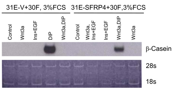
Figure 7
The Wnt-3a-imposed abrogation of differentiated response to lactogenic hormones was mitigated in pBabeSFRP-4-infected Line 31E cells. Using Northern blot methods, the expression of β-casein was investigated after stimulation of cultures with Wnt-3a and/or different combinations of growth factors and lactogenic hormones. Lines 31E +30F co-cultures containing either pBabeVector or pBabeSFRP-4-infected Line 31E cells were compared. Most notable is that Wnt-3a is capable of completely suppressing the differentiation to β-casein expression stimulated by dexamethazone-insulin-prolactin (DIP). However, co-cultures containing Line 31E cells infected with SFRP-4 show definite β-casein induction in spite of Wnt-3a treatment. Ribosomal RNA was stained with acridine orange as a loading control.