
Figure 1
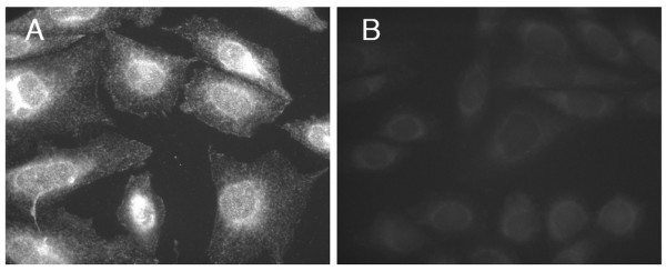
Preadsorbtion control showing the specificity of the Anti-sigma-1 receptor antibody. CHO-K1 cells stained with anti-sigma1 antibody (0.6 μg/ml). A – no preadsorbtion; B – before immunostaining antibody was preadsorbed for 1 h with the purified sigma-1 protein (25 μg/ml).
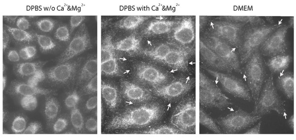
Figure 2
In CHO-K1 cells during a 30 sec rinsing with buffers with or w/o Ca2+ and Mg2+ before fixation changed the intracellular distribution of endogenous sigma-1 receptors. A. Cells were rinsed with DPBS w/o Ca2+ and Mg2+ and DMEM. B. Cells were rinsed with DPBS with different concentrations of Ca2+ and Mg2+
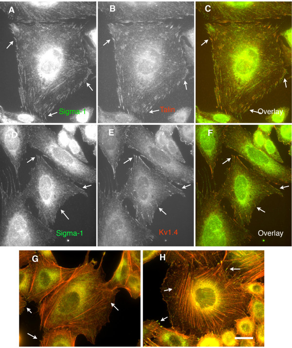
Figure 3
Endogenous Sigma-1 receptors colocalize with other proteins in the FAC. A-C. Colocalization with talin. A-sigma-1; B-talin; C-Overlay. D-F. Colocalization with Kv1.4. potassium channel. D-sigma-1; E-Kv1.4.; F-Overlay. G, H. Colocalization of sigma-1(green) and F-Actin(red) in mock or phalloidin (50 ng/ml for 30 min) treated cells. Note, that the sigma-1 localized on the the barbed ends of actin filaments.
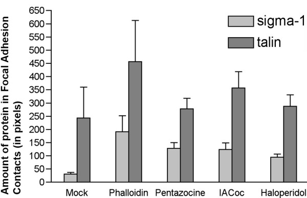
Figure 4
Dynamics of endogenous Sigma-1 receptor and Talin appearence in the FAC in CHO-K1 cells under various treatment conditions. All the compounds indicated were added to cells in complete 10% FBS supplemented DMEM for 30 min at a concentration of 10 μM except phalloidin (50 nM). Morphometrical quantitation of the amount of the sigma-1 receptor or talin in the FAC was performed with the Axiovision 4.3 software. The total amount of staining (in pixels) for each channel was quantitated for each image and divided by the number of cells (detected by DAPI staining of nuclei). 101–155 cells were measured per each condition.
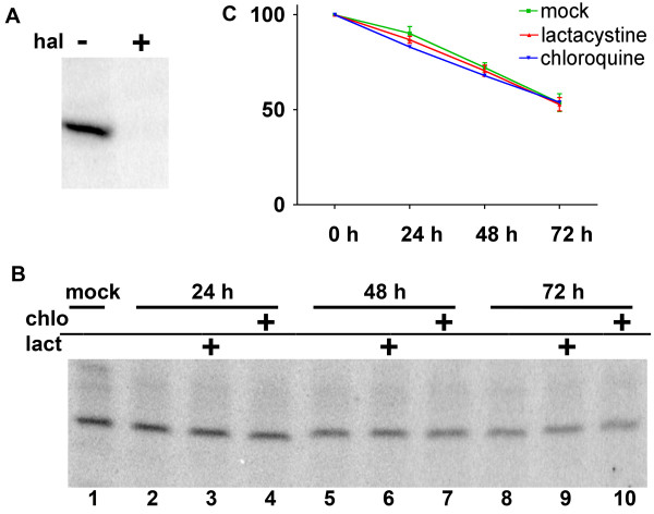
Figure 5
Stability of the sigma-1 receptors in CHO-K1 cells. Endogenous sigma-1 receptors were [125I]-IACoc photolabeled in live CHO-K1 cells by a brief photolysis (see Materials and Methods for details). Labeled cells were washed free of excess ligand and plated on culture dishes for various periods of time with or without inhibitors of proteolysis. A. Specificity of I125-azidococaine labeling in live cells was tested by preincubation of cells with 5 μM haloperidol (hal), prior to [125I]-IACoc labeling (plus conditions). B. Autoradiogram of CHO-K1 cell lysate collected at different time points after plating of photolabeled cells: time 0 – (lane # 1); after 24 h – (lanes # 2,3,4); after 48 h – (lanes # 5,6,7); after 72 h – (lanes # 8,9,10). The proteosomal inhibitor, lactacystine, 2 μM (lanes #3,6,9) was added; the lysosomal inhibitor, chloroquine, 100 nM (lanes # 4,7,10) was added. C. Quantified data from the situ photolabeled sigma-1 receptor time-course from 3 independent experiments are represented in part B. These data indicate that in CHO-K1 cells the turnover of the ligand occupied sigma-1 receptor is slow with a half-life at least 72 hours.