
Figure 1
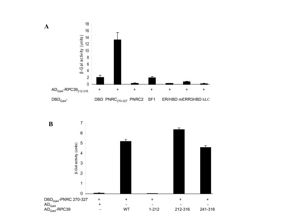
PNRC interacts with a subunit of RNA pol III. A, ADGal4-RPC39212–316 expression plasmid was isolated from a human mammary gland expression library screening using DBDGal4-PNRC270–327 as bait. To confirm the interaction and the specificity of the interaction between PNRC and RPC39, yeast strain Y187 was cotransformed with the expression plasmids for the expression of the fusion proteins as indicated, and transformants containing these plasmids were selected by growth on SD/-Leu/-Trp agar plates. The expression of interacting hybrid proteins in Y187 transformants was analyzed for LacZ expression. DBDGal4 (DBD) and DBDGal4-human lamin C (hLC) expression plasmids were included as background and negative control, respectively. β-Galactosidase activities in liquid cultures are expressed in Miller units as mean ± s.d. of three independent assays. B, PNRC interacts with the C-terminus, amino acids 212–316, of the human Pol III subunit RPC39. The yeast expression plasmids, pACT2-RPC39 and pACT2-RPC391–212, for the expression of ADGal4-RPC39 wild type (WT) or ADGal4-RPC1–212 (1–212) fusion proteins were constructed as described in 'Methods'. The expression plasmids for ADGal4-RPC39212–316 (212–316) and ADGal4-RPC39241–316(241–316) were isolated from library screening through their interaction with PNRC270–327. Y187 cells were cotransformed with the expression plasmids coding for the fusion proteins as indicated. The selection of Y187 transformants and the β-Gal assays on transformants were performed as described in A.
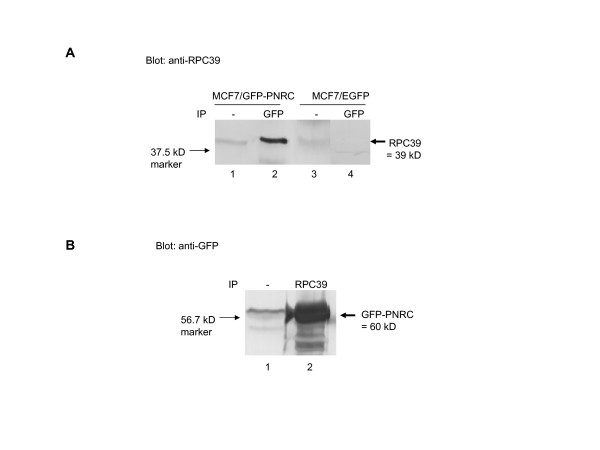
Figure 2
PNRC associates with RPC39 in mammalian cells. MCF-7/EGFP or MCF7/EGFP-PNRC stable expression cells were transiently transfected with the RPC39 expression plasmid, pSG5-RPC39, 24 h post-transfection, cells were harvasted and lysed, and 15 mg of total proteins were immunoprecipitated with antibodies against GFP (Clontech) or RPC39 (Santa Cruz Biotechnology). Specifically bound proteins to Protein A agarose beads were separated on a 10% SDS-PAGE and analysed by immunoblotting with either anti-RPC39 (A, blot: anti-RPC39) or anti-GFP (B, blot: anti-GFP) antibody, as previously described [2]. An aliquot of cell lysate equal to 100 μg of protein was included in each SDS-PAGE gel for Western blot to examine whether the crude protein extracts used for co-immunoprecipitation contains EGFP-PNRC and RPC39 proteins. The protein bands with molecular weights corresponding to those of RPC39 or EGFP-PNRC were indicated by arrows.
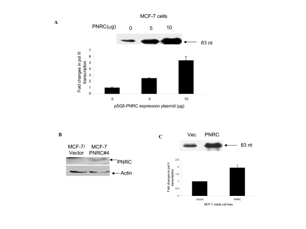
Figure 3
PNRC stimulates the transcription of tRNAarg gene by Pol III. MCF7 breast cancer cells (A) were transiently transfected with 10 μg of reporter plasmid pArg-maxi which contains a Drosophila tRNAarg gene and increasing amounts (0, 5, or 10 μg) of PNRC expression plasmid, pSG5-PNRC. Cells were cultured for 24 hours and the tRNAarg transcripts in 1 μg of total RNA isolated from the transfected cells was determined by RNase protection assay using a 32P-labeled antisense riboprobe. An example of an autoradiogram of tRNAarg transcript (indicated by an arrow) and quantification of the levels of tRNAarg mRNA from three independent experiments are shown. B, Western blot analysis of PNRC protein in MCF7/vector and MCF7/PNRC stably transfected cells. The procedures for transfection, G418 selection, and individual clone screening were described in Methods. An aliquot of cell lysate, from MCF7/vector or MCF7/PNRC cells, equal to 100 μg of protein was analysed by Western blot using PNRC antiserum (1:500 dilution) or anti-actin antibody (1:1000 dilution) as previously described [2]. The protein bands with molecular weights corresponding to those of PNRC or actin were indicated by arrows. C, MCF-7/vector or MCF7/PNRC stable expression cells were transiently transfected with 10 μg of reporter plasmid pArg-maxi. Twenty-four hours after transfection, the tRNAarg mRNA levels in the transfected cells were analysed by RNase protection assay as described for A. An example of an autoradiogram of tRNAarg transcript (indicated by an arrow) and quantification of the levels of tRNAarg mRNA from three independent experiments are shown.
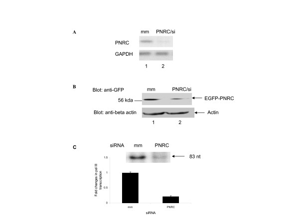
Figure 4
Down regulation of RNA Pol III gene transcription by specific depletion of endogenous PNRC using RNA interference. A, Northern blot analysis of PNRC and GAPDH mRNAs. MCF7 cells were transiently transfected with Pol III reporter plasmid, pArg maxi, along with 20 nM of either siRNA specific for PNRC (PNRC/si) or nonspecific mismatch RNA (mm). Twenty-four hours after transfection, 20 μg of total RNA, isolated from the transfected cells, was subjected to Northern analysis with PNRC and GAPDH cDNA probes, separately, as described in Methods section. B, Western blot of PNRC and actin protein. An aliquot of cell lysate equal to 100 μg of protein prepared from PNRC/siRNA (20 nM) transfected (PNRC/si) or nonspecific mismatch RNA (mm) transfected MCF7/EGFP-PNRC cells was separated on a 10% SDS-PAGE and subjected to Western analysis with 1:1000 diluted anti-GFP mouse monoclonal antibody (Cloetech) and anti-actin antibody (Santa Cruz Biotechnology) separately. The protein bands with molecular weights corresponding to those of EGFP-PNRC fusion protein or actin were indicated by arrows. C, Down regulation of tRNAarg transcription by PNRC/siRNA. MCF-7 cells were transiently transfected with reporter plasmid, pArg maxi (10 μg) along with 20 nM of either PNRC/siRNA (PNRC) or mismatch control RNA (mm). Twenty-four hours after transfection, the levels of tRNA transcripts in the transfected cells were determined by RNase protection assay as described in figure 3.
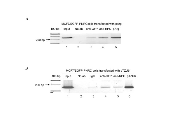
Figure 5
Co-recruitment of PNRC and RPC39 onto RNA polymerase III-dependent genes. MCF-7/EGFP-PNRC cells were transfected either with pArg maxi reporter plasmid, which contains tRNAarg gene promoter and coding sequence, or with pTZU6 plasmid that carries the U6 RNA gene promoter. Twenty-four hours after transfection, cells were treated with formaldehyde to crosslink endogenous proteins and DNA. Samples of sonicated and purified chromatin were immunoprecipitated with no antibody (No ab), preimmuno IgG (IgG), GFP antibody (anti-GFP), or RPC39 antibody (anti-RPC) as indicated. DNA isolated from immunoprecipitated material was amplified by PCR with primers to amplify a 220 bp fragment of the Drosophila tRNAarg gene (A) or a 160 bp fragment of the U6 RNA gene (B). The amplified PCR products were analyzed on 1.8% agarose gel. The PCR products from the reactions using input DNA, pArg or pTZU6 plasmids as template were included on the gels as positive and size controls.