
Figure 1
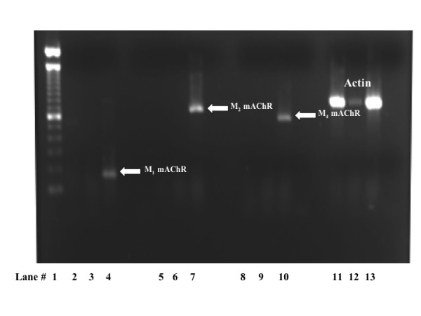
Mouse embryonic fibroblasts (MEF) cells do not express mRNA encoding M1, M2 or M4 mAChR subtypes. A representative gel showing lack of mAChR expression in MEF wild type and KO1/2 cells Lanes consisted of 100 bp ladder (lane 1), wild type MEF cells (lanes 2, 5, 8 and 11), MEF β-arrestin KO1/2 (lanes 3, 6, 9, and 12), rat cortex (lanes 4 and 10 and 13), and rat cerebellum (lane 7). The white arrows point to M1, M2 and M4 mAChR PCR product from cDNA as positive controls. The electrophoresis gel shown is a representative of at least 3 independent experiments.
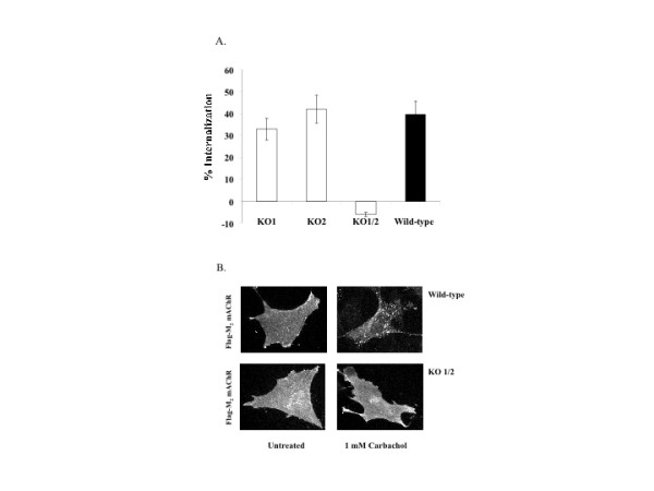
Figure 2
Agonist-promoted internalization of M2 mAChR in MEFs is β-arrestin-dependent. A.) Approximately 24 h following transfection with M2 mAChR, cells were stimulated with 1 mM carbachol for 1 h and agonist-promoted internalization was determined using [3H-NMS]. Data are presented as the mean ± standard error from 3 separate experiments with each experiment consisting of 8 to 11 independent determinations. Statistical test was performed using ANOVA with the post hoc Bonferroni/Dunn test (asterisk indicates * p < 0.001). B.) Cells were transfected as described above and then incubated in the presence or absence of 1 mM carbachol for 30 minutes prior to indirect immunofluorescence as described in Methods. Images were acquired at 40X.
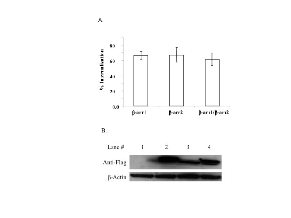
Figure 3
Expression of β-arrestin 1 or 2 rescued agonist-promoted internalization of M2 mAChRs in MEF KO1/2 cells. Approximately 24 h following co-transfection with constructs encoding M2 mAChRs and β-arrestins, cells were stimulated with 1 mM carbachol for 1 h. A.) Agonist-promoted internalization was determined as described in Methods. Data are presented as the mean ± standard deviation of 5 independent experiments consisting of 8–11 determinants. B.) A representative immunoblot of FLAG-tagged β-arrestin and internal protein control β-actin is shown. Lanes consisted of: non-transfected MEF KO1/2 (1), MEF KO1/2 expressing β-arrestin1 (2), MEF KO1/2 expressing β-arrestin 2 (3), and MEF KO1/2 expressing β-arrestin 1 and 2 (4). Western blot shown is a representative of at least 3 independent experiments.
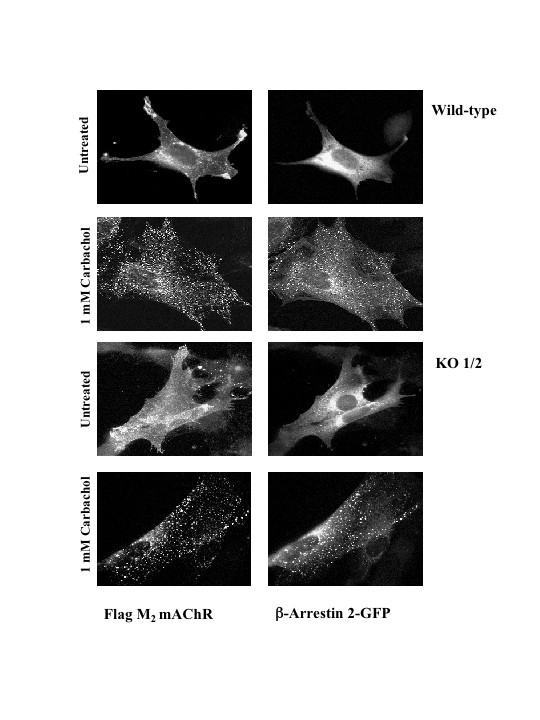
Figure 4
Stimulation of M2 mAChRs leads to stable co-localization of β-arrestin 2-GFP at intracellular sites. MEF wild type or KO1/2 cells were transiently co-transfected with the human FLAG-tagged M2 mAChR and β-arrestin 2-GFP constructs. Following 30 minutes of 1 mM carbachol stimulation, cells were fixed and processed for indirect immunofluorescence as described in the Methods. Localization of β-arrestin 2-GFP and M2 mAChR was visualized by confocal microscopy. Confocal images are representative of three independent experiments.
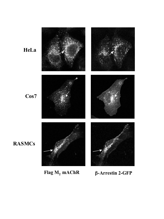
Figure 5
Agonist-promoted internalized FLAG-tagged M2 mAChRs exhibit overlap with β-arrestin 2-GFP at intracellular sites in various cell lines. Cells were transiently transfected with FLAG-tagged M2 mAChR and β-arrestin 2-GFP and treated with 1 mM carbachol for 30 min at 37°C. Co-localization of β-arrestin 2-GFP with internalized M2 mAChRs occurred in HeLa, COS-7 and rat aortic smooth muscle cells (RASMCs). Arrows indicate overlap between β-arrestin 2-GFP and M2 mAChRs in intracellular compartments. Confocal images are representative of three independent experiments.
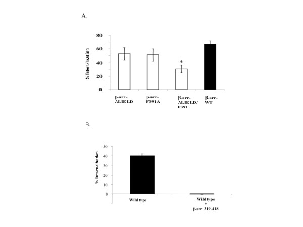
Figure 6
Expression of β-arrestin mutants deficient in clathrin and/or AP-2 binding interaction partially supports agonist-promoted internalization of M2 mAChRs in MEF K/O1/2, while expression of truncated carboxyl-terminal region of β-arrestin 1 (319–418) completely blocked agonist promoted M2 mAChR internalization in MEFwt cells. A.) Approximately 24 hr following co-transfection with FLAG-M2 mAChR and β-arrestin 2 clathrin (ΔLIELD), AP-2 (F391A), or clathrin and AP-2 (ΔLIELD/F391A) mutants, MEF KO1/2 cells were stimulated with 1 mM carbachol for 1 h and agonist-promoted internalization was determined as described in Methods. Data are presented as mean ± standard deviation from 4 independent experiments consisting of 8–11 determinants. B.) Approximately 24 hr following transfection with the β-arrestin 1 C-terminal domain (319–418), MEF wild type cells were stimulated with 1 mM carbachol for 1 h and agonist-promoted internalization of receptor was determined as described in Methods. Data are presented as the mean ± standard error from 3 separate experiments with each experiment consisting of 8 to 11 independent determinations. Statistical test was performed using ANOVA with the post hoc Bonferroni/Dunn test (asterisk indicates * p < 0.001).
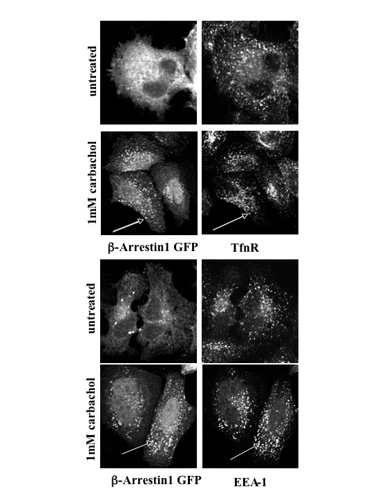
Figure 7
Addition of agonist leads to the redistribution of β-arrestin 1-GFP to early endosomal structures in the cytosol. HeLa cells were transiently transfected with human FLAG-tagged M2 mAChR and β-arrestin 1-GFP and treated with 1 mM carbachol for 30 minutes. Cells were processed for confocal microscopy. β-arrestin 1-GFP complexes localized to the early endosome as shown by colocalization with markers of that compartment (EEA-1 and TfnR). Arrows indicate signficant overlap between TfnR or EEA-1 with β-arrestin 1-GFP. Confocal images are representative of three independent experiments.
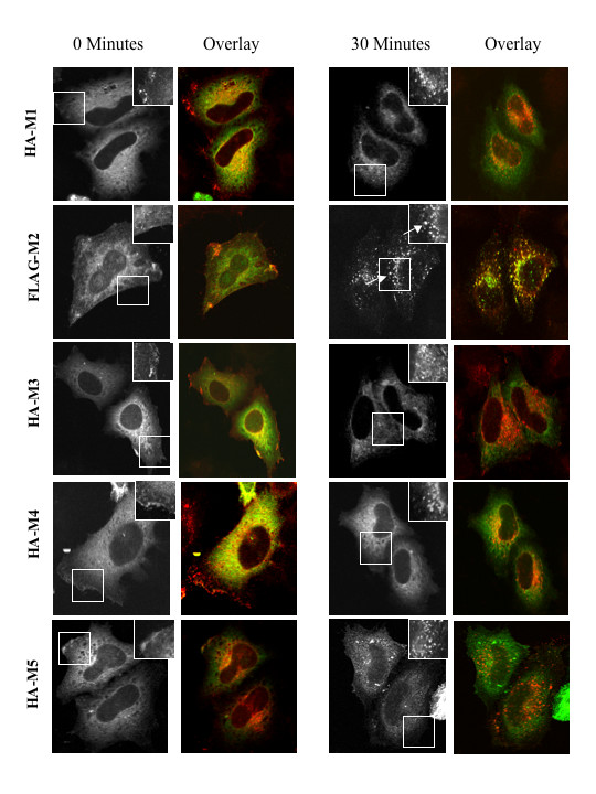
Figure 8
Internalized M2 mAChRs exhibit a differential affinity for β-arrestin 2-GFP compared to other muscarinic subtypes. HeLa cells were transiently co-transfected with plasmids encoding β-arrestin 2-GFP and either HA-tagged M1, M3, M4, M5 mAChR or FLAG-tagged M2 mAChR. Cells were untreated or treated with 1 mM carbachol for 0 min or 30 min. Grayscale image indicates β-arrestin 2-GFP localization while the upper right inset indicates immunostaining of the mAChR in a small section of the cell (outline). Arrows indicate overlap between internalized M2 mAChRs and β-arrestin 2-GFP. Overlay represents co-immunostaining of mAChR (red) and β-arrestin 2-GFP expression (green) and their colocalization (yellow).