
Figure 1
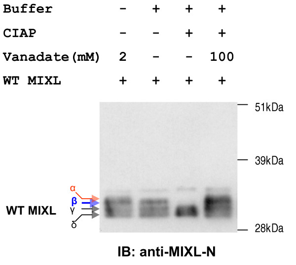
Alkaline Phosphatase treatment of nuclear extracts alters the mobility of MIXL1 proteins on SDS-PAGE gel. Expression plasmid pCMV5-MIXL1 was transiently transfected to HEK 293T cells and the transfectants were harvested 60 hours later. 15ug of fresh nuclear extracts were treated with 40u calf intestine alkaline phosphatase (CIAP) in the absence or presence of phosphatase inhibitor sodium orthovanadate at 30°C for 15 min. After CIAP treatment, nuclear extracts with orthovanadate and CIAP reactions were resolved on a NuPAGE gel and transferred to a PVDF membrane. Immunobloting was done with anti-MIXL1-N (1:350 dilution). Anti-MIXL1-N detected 4 species on lane 1 (nuclear extracts with orthovanadate and no CIAP treatment). The band α disappeared on mock reaction (lane 2). CIAP treatment resulted in disappearance of both band α and β (lane 3). In contrast, the addition of phosphatase inhibitor orthovanadate protected the all the 4 species (lane 4).
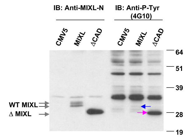
Figure 2
MIXL1 is phosphorylated on tyrosine residues. Nuclear proteins were extracted from 293T cells transiently transfected with constructs pCMV5-MIXL1 and pCMV5-ΔCAD. Equal amounts of nuclear extracts (10 μg for CMV5 control and MIXL1 and 5 μg for ΔCAD) were loaded in duplicate into a NuPAGE gel and transferred to PVDF membranes. The blot was divided into two halves and hybridized with either anti-MIXL1-N (1:100) or anti-P-Tyr (mouse monoclonal antibody 4G10 at 1:4000). Anti-MIXL1-N antibody detected two species in transfectants expressing full-length MIXL1 and a strong fuzzy band in transfectants expressing the C-terminal truncation. The second half of the blot probed with 4G10 detected a weak but specific signal (arrow) corresponding to the slower migrating form of full-length MIXL1 and a robust signal (arrow) for the C-terminal truncation.
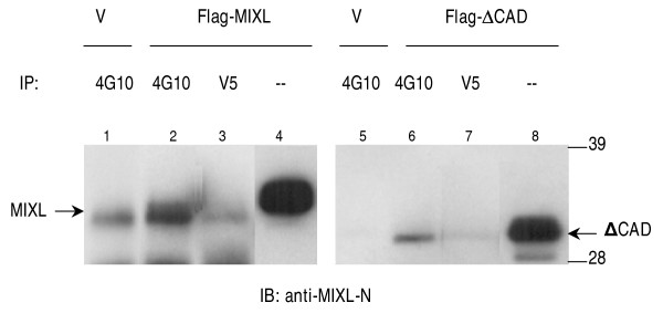
Figure 3
Immunoprecipitation of phosphorylated MIXL1 by anti-phosphotyrosine antibody 4G10. Expression constructs pCMV2-flag-MIXL1 and pCMV2-flag-ΔCAD were transfected into HEK 293T cells. 500 μL of nuclear proteins (1μg/μL) from the transfected cells were precleared with 50 μL protein-A-agarose bead slurry (50%v/v) and incubated with 5 μg of the phosphotyrosine-specific antibody 4G10 or the antibody anti-V5 as an isotypic control overnight. The immune complexes were precipitated with 60 μL protein-A-agarose bead slurry (50%v/v). Washed pellets were dissociated and resolved in a NuPAGE gel for immunobloting with the antibody anti-MIXL1-N (1:500). The immunoprecipitation and immunobloting for full-length MIXL1 is shown in the left panel, while mutant ΔCAD in the right panel. A small fraction of both full-length MIXL1 and mutant ΔCAD were immunoprecipitated with the antibody 4G10 (Lane 2 or 6) but not with an isotypic control antibody anti-V5 (Lane 3, or 7). Supernatants from the immunoprecipitation reactions denoted by (-) show the integrity of proteins. A strong band below full-length MIXL1 appeared to be non-specific, as it was also present in the controls. The blot on the left (lanes 1, 2, 3) was exposed to a Kodak film for 30 seconds, while the blot on the right (lanes 4, 5, 6) was exposed for only 5 seconds.
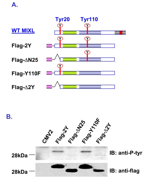
Figure 4
MIXL1 is phosphorylated on Tyr20. A) Cartoon depicting MIXL1 constructs generated for mutational analysis. Three MIXL1 mutant constructs were derived from the ΔCAD mutant construct pCMV2-flag-ΔCAD (renamed as Flag-2Y in this figure). Each mutant in the constructs has a Flag epitope (a solid gray bar) attached in frame to the amino-terminus and contains either tyr20 or tyr110 or none as illustrated. B) Detection of tyrosine phosphorylation. The mutant constructs were transiently transfected into HEK 293T cells. 10 μg of nuclear proteins from the transfectants were resolved on a NuPAGE gel. Tyrosine phosphorylation was examined by immunobloting with monoclonal antibody 4G10 (1:3000). The blot was stripped and re-probed with mouse monoclonal antibody anti-flag (1:300). Tyrosine phosphorylation was detected in both the mutant Flag-2Y and the mutant Flag-Y110F but not in the mutant Flag-ΔN25 as well as the mutant Flag-Δ2Y. The results are consistent with Tyr20 being the target of phosphorylation.
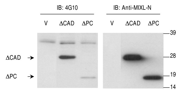
Figure 5
Tyr20 phosphorylation is diminished in the absence of PRD. The constructs pCMV5-ΔCAD and pCMV5-ΔPC (lacking both the CAD and PRD) were transfected into HEK 293T cells. 15 μg of the nuclear extracts from the transfectants were resolved on a 10% NuPAGE gel. Tyrosine phosphorylation was detected by immunobloting with monoclonal antibody 4G10. The same blot was stripped and reprobed with anti-MIXL1-N antibody (1:400). Compared to the vector control, the antibody 4G10 detected a weak signal for the ΔPC mutant, although it is much weaker than that for ΔCAD mutant with the intact PRD domain. V-Vector.