
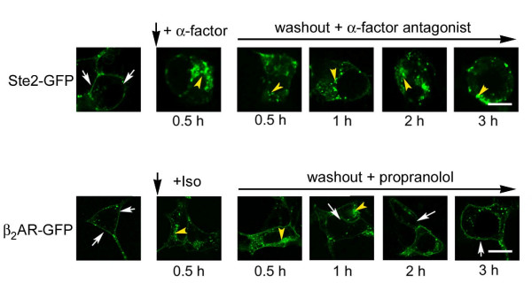
Figure 1
Trafficking of yeast Ste2 in human HEK293 cells in response to α-factor: analysis by confocal microscopy. HEK cells were transiently transfected to express either Ste2-GFP or β2AR-GFP. Unstimulated cells display receptors that are largely localized to the cell membrane (white arrows denote cell membrane-localized receptors). Cells expressing yeast Ste2-GFP and challenged with α-factor (10 μM, upper panel) as well as those expressing β2AR-GFP and challenged with beta-adrenergic agonist isoproterenol (Iso, 10 μM; lower panel) for 0.5 hr displayed frank internalization (yellow arrowheads denote internalized receptors). Agonist ligands were then washed from the media and the cells treated the appropriate antagonist ligand. Possible recycling of internalized receptors back to the cell membrane was followed for 3 hours. For cells expressing Ste2-GFP, the a-factor antagonist (des-Trp, Ala-3 analog of a-factor, 10 μM) was added after wash-out and the cells were monitored at 0.5 (panel c), 1 (panel d), 2 (panel e), and 3 (panel f) hour time periods after wash-out of agonist. For cells expressing β2AR-GFP, the β-adrenergic antagonist propranolol (10 μM) was added after wash-out of isoproterenol and the cells were monitored at 0.5 (panel i), 1 (panel j), 2 (panel k), and 3 (panel l) hour time periods post wash-out of agonist. The images displayed are from a single experiment, representative of more than five replicate, separate experiments. Bar equals 10 μm.
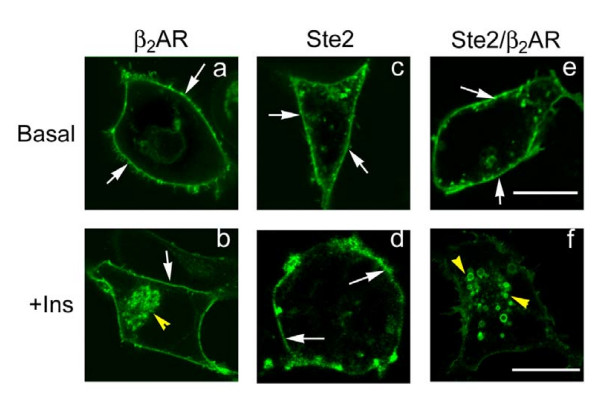
Figure 2
Trafficking of yeast Ste2, human β2AR, and a yeast Ste2/human β 2AR chimera expressed in human HEK293 cells in response stimulation by insulin: analysis by confocal microscopy. HEK293 cells were transiently transfected to express either β2AR-GFP (panels a, b), Ste2-GFP (panels c, d), or the Ste2/β2AR-GFP chimeric receptor (panels e,f). For each of these GPCRs, localization of the receptors in the untreated cells was dominant at the cell membrane (white arrows). Cells were treated without (panels a, c, e) or with (panels b, d, f) insulin (100 nM) for 0.5 h and followed by confocal microscopy. Insulin stimulates internalization of the β2AR-GFP (panel b) and of the Ste2/β2AR-GFP chimeric receptor (panel f), but not of Ste2-GFP (panel d). The images displayed are from a single experiment, representative of more than five replicate, separate experiments. Bar equals 10 μm.